Irene Faure de Pebeyre
|
|
| email |
|
|
| phone & fax |
Chirurgische Klinik und Poliklinik Innenstadt
NARVIS lab
Phone: +49 (89) 5160-3615
Fax: +49 (89) 5160-3630
|
| address |
Irene Faure de Pebeyre
Institut für Informatik I-16
Technische Universität München,
Boltzmannstr. 3
85748 Garching b. München
Germany
|
|

|
Diploma Thesis
Publications
Research Projects
|
|
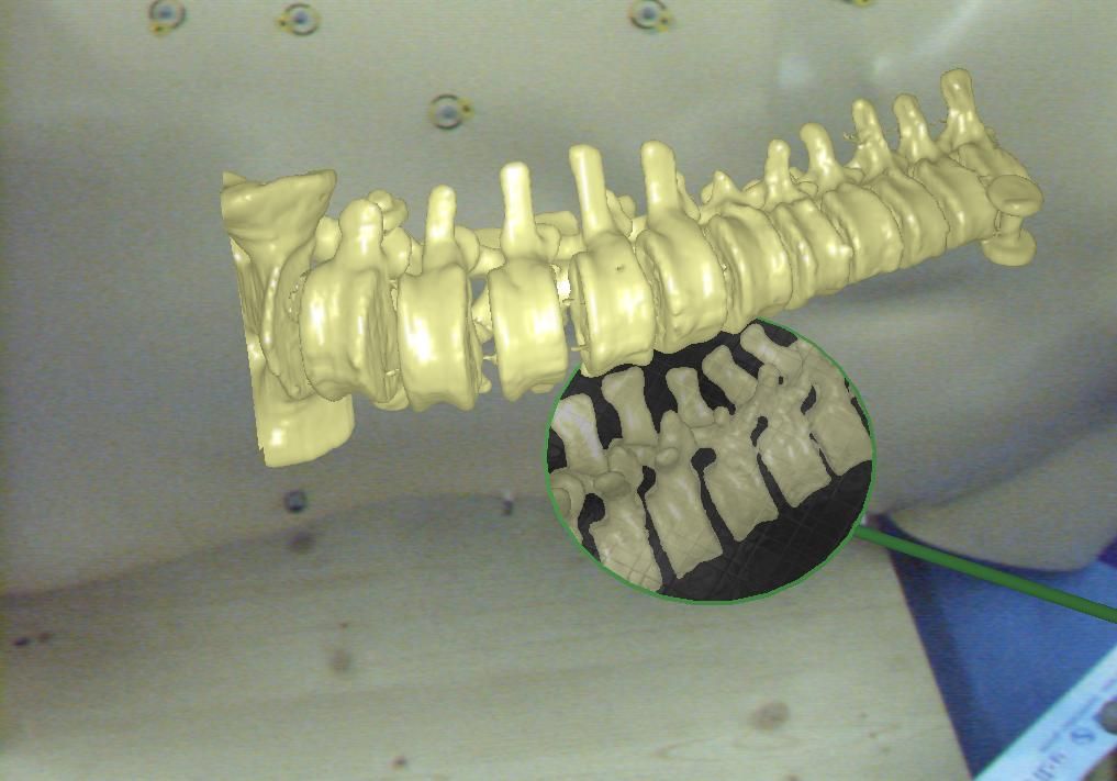
Augmented Reality offers a higher degree of freedom for the programmer than classical visualization of volume data on a screen. The existing paradigms for interaction with 3D objects are not satisfactory for particular applications since the majority of them rotate and move the object of interest. The classic manipulation of virtual objects cannot be used while keeping real and virtual spaces in alignment within an AR environment. This project introduces a simple and efficient interaction paradigm allowing the users to interact with 3D objects and visualize them from arbitrary viewpoints without disturbing the in-situ visualization, or requiring the user to change the viewpoint. We present a virtual, tangible mirror as a new paradigm for interaction with 3D models. The concept borrows its visualization paradigm in some sense from methodology used by dentists to examine the oral cavity without constantly changing their own viewpoint or moving the patients head. The virtual mirror improves the understanding of complex structures, enables completely new concepts to support navigational aid for different tasks and provides the user with intuitive views on physically restricted areas.
|
|
|
Intra-operative localization of non-superficial cancerous lesions in non-hollow organs like liver, kidney, etc is currently facilitated by intra-operative ultrasound (IOUS) and palpation. This yields a high rate of false positives due to benign abnormal regions and thus unnecessary resections with increased complications and morbidity. In this project we integrate functional nuclear information from gamma probes with IOUS, to provide a synchronized, real-time visualization that facilitates the detection of active tumors and metastases intra-operatively. The bet of this project is that the inclusion of an advanced, augmented visualization provides more reliability and confidence on classifying lesions prior to the resection.
|
|
|
Nuclear medicine imaging modalities assist commonly in surgical guidance given their functional nature. However, when used in the operating room they present limitations. Pre-operative tomographic 3D imaging can only serve as a vague guidance intra-operatively, due to movement, deformation and changes in anatomy since the time of imaging, while standard intra-operative nuclear measurements are limited to 1D or (in some cases) 2D images with no depth information. To resolve this problem we propose the synchronized acquisition of position, orientation and readings of gamma probes intra-operatively to reconstruct a 3D activity volume. In contrast to conventional emission tomography, here, in a first proof-of-concept, the reconstruction succeeds without requiring symmetry in the positions and angles of acquisition, which allows greater flexibility and thus opens doors towards 3D intra-operative nuclear imaging.
|
|
|
In minimally invasive tumor resection, the goal is to perform a minimal but complete removal of cancerous cells. In the last decades interventional beta probes supported the detection of remaining tumor cells. However, scanning the patient with an intraoperative probe and applying the treatment are not done simultaneously. The main contribution of this work is to extend the one dimensional signal of a nuclear probe to a four dimensional signal including the spatial information of the distal end of the probe. This signal can be then used to guide the surgeon in the resection of residual tissue and thus increase its spatial accuracy while allowing minimal impact on the patient.
|


