Past Projects
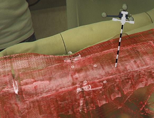
ARAV Augmented Reality Aided VertebroplastyIn today’s ORs more and more operations are performed employing minimally invasive procedures. Surgical instruments are inserted through a tiny cut on the patient’s skin, the port to the inside of the patient. In some cases endoscope cameras record video images of the operation site that are presented on a monitor. As a consequence of this technique, the surgeon’s field of view is divided into several work spaces, the monitor, the patient and information of medical imaging data presented on a third station. The missing direct view on the workspace complicates intuitive control of surgical tools. In contrast to open surgery the physician has to collect information from several fields of view at the same time and fuse information mentally to create a complete model of his working space, the operation site. The minimally invasive intervention vertebroplasty was determined as a suitable medical application to bring an Head Mounted Display (HMD) into the OR for augmentation of surgical instruments and medical imaging data. In-situ visualization with an HMD presents all available imaging data and navigational information in one field of view. The objective of vertebroplasty is the insertion of cement into weak and brittle vertebrae through a trocar for stabilization. In this case the view on the inside of the patient is not provided by an endoscope camera. However, since the operation is performed under a CT scanner, imaging data is permanently updated to check position of the trocar and amount of inserted cement. Imaging data is presented on a monitor and has to be mapped mentally by the surgeon on the real operation site. |
Surgical Workflow Analysis in Laparoscopy for Monitoring and DocumentationSurgical workflow recovery is a crucial step towards the development of intelligent support systems in surgical environments. The objective of the project is to create a system which is able to recognize automatically the current steps of a surgical laparoscopic procedure using a set of signals recorded from the OR. The project adresses several issues such as the simultaneous recordings of various signals within the OR, the design of methods and algorithms for processing and interpreting the information, and finally the development of a convenient user interface to display context sensitive information inside the OR. The current clinical focus is on laparoscopic cholecystectomies but the concepts developed in the project also apply to laparoscopic surgeries of other kinds. |
Reconstruction and Registration of Histology and Phase Contrast Images for Clinical Validation of Imaging ModalitiesBefore its introduction into the hospital, a new imaging modality has to be validated. In other words, appearing structures need to be correlated to the imaged tissues. Such a cross validation is only meaningful when performed against the gold standard which is histology. Currently, cross validation is performed by qualitative comparison of 3D datasets to 2D histology slices. Since the acquisition of a consistent 3D histology volume is a challenging task, its comparison to the corresponding dataset always remained qualitative.The classic histology procedure can be divided in four steps: pre processing, cutting, post processing and imaging of the tissues. In the first step, the sample is chemically processed to preserve the tissues and is then embedded in a paraffin block. By using a microtome, it is cut in very thin slices, and put on a glass slide. During post processing the sample is stained to enhance the structures of interest. The imaging is performed with a camera mounted in a microscope, or with a dedicated scanner. Several difficulties inherent to this process can have a dramatic influence on the quality of the reconstructed histology volume. For instance, since the cutting process is done manually, problems like flipping, bending or ripping of the slides may happen. If the knife starts to wear off, some banding will appear over the slices. Moreover, a few slices could be missing. Finally since the staining color is time dependent, variation in the color of the slices can occur. In this project, we propose to improve the histology procedure and to develop methods towards a consistent reconstruction of 3D histology volumes. |
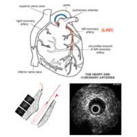
Assessment of Fluid Tissue Interaction Using Multi-Modal Image Fusion for Characterization and Progression of Coronary AtherosclerosisCoronary artery diseases such as atherosclerosis are the leading cause of death in the industrialized world. In this project, we develop computational tools for segmentation and registration problems on intravascular images including IVUS (Intravascular Ultrasound) and OCT (Optical Coherence Tomography). One sample component of this project is Automatic Stent Implant Follow-up from Intravascular OCT Pullbacks. The stents are automatically detected and their distribution is analyzed for monitoring of the stents: their malpositioning and/or tissue growth over stent struts. |
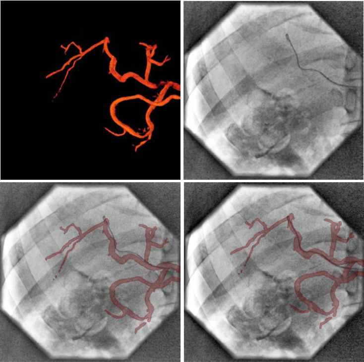
Motion Compensation for CatheterizationsIn many minimally-invasive interventions, catheters are inserted into the body and guided to a region of interest with the help of fluoroscopic imaging (low-dose X-ray image sequences). Due to patient motion, the navigation can be disturbed, and the physician needs a longer time for treatment. This is hazardous in terms of radiation for both, physician and patient. In order to overcome these problems, suitable image-based compensation methods to resolve (non-) rigid motion can be applied. In this project, patient motion is analyzed and suitable compensation algorithms are developed. |
MeTaTop A Multi Sensory Table Top System for Medical ProceduresA tabletop system in medical environments can be used for interactive and collaborative analysis of patient data but also as a multimedia user interface within sterile space. For preoperative planning physicians in charge with a particular patient meet to discuss the medical case and plan further steps for therapy. For this reason, they could collaboratively view and browse through all kind of available medical imaging data with the tabletop system. Alternatively such a system could be a central interaction device for all kind of equippment within the OR requiring user input, however, can not be operated by the sterile surgeon. We believe that the projection of all kind of user and information interfaces on a sterile glass plane would facilitate the clinical workflow.This project is strongly related to the Tangible Interaction Surface for Collaboration between Humans project. |
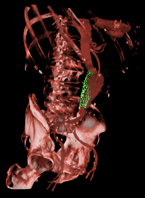
Endovascular Stenting of Aortic AneurysmsEndovascular stenting is a minimally invasive treatment technique for aortic aneurysms or dissections. Thereby, a certain aortic prosthesis (stent graft) is placed inside the aortic aneurysm in order to prevent a life-threatening rupture of the aortic wall. Prior to the intervention, a computed tomography angiography (CTA) is acquired on which the surgical staff can measure the parameter of the desired stent graft and finalize the intervention workflow. The entire interventional catheter navigation is done under 2D angiography imaging where the physician is missing the important 3D information. The purpose of our project is two-fold:1. In the planning phase, a modified graph cuts algorithm automatically segments the aorta and aneurysm, so the surgical staff can choose an appropriate type of stent to match the segmented location, length, and diameter of the aneurysm and aorta. By visualizing the defined stent graft next to the three-dimensionally reconstructed aneurysm, mismeasurements can be detected in an early stage. Our main goal is the creation of an interactive simulation system that predicts the behaviour of the aortic wall and the movement of the implanted stent graft. 2. During implantation of the stent graft, after an intensity based registration of CTA and angiography data, the current navigation can be visualized in the 3D CT data set at any time. This includes solutions for electro-magnetic tracking of catheters as well as guide wires and stent grafts. Eventually, Our main goal is the creation of solutions that enable the surgeon to enhance the accuracy of the navigation and positioning, along with a minimum use of angiography, leading to less radiation exposure and less contrast agent injection. |
Port Placement in Minimally Invasive Endoscopic SurgeryOptimal port placement is a delicate issue in minimally invasive endoscopic surgery. A good choice of the instruments' and endoscope's ports can avoid time-consuming consecutive new port placement. We present a novel method to intuitively and precisely plan the port placement. The patient is registered to its pre-operative CT by just moving the endoscope around fiducials, which are attached to the patient's thorax and are visible in its CT. Their 3D positions are automatically reconstructed. Without prior time-consuming segmentation, the pre-operative CT volume is directly rendered with respect to the endoscope or instruments. This enables the simulation of a camera flight through the patient's interior along the instruments' axes to easily validate possible ports. |
Laparoscope Augmentation for Minimally Invasive Liver ResectionIn recent years, an increasing number of liver tumor indications were treated by minimally invasive laparoscopic resection. Besides the restricted view, a major issue in laparoscopic liver resection is the precise localization of the vessels to be divided. To navigate the surgeon to these vessels, pre-operative imaging data can hardly be used due to intra-operative organ deformations caused by appliance of carbon dioxide pneumoperitoneum and respiratory motion.Therefore, we propose to use an optically tracked mobile C-arm providing cone-beam computed tomography imaging capability intra-operatively. After patient positioning, port placement, and carbon dioxide insufflation, the liver vessels are contrasted and a 3D volume is reconstructed during patient exhalation. Without any further need for patient registration, the volume can be directly augmented on the live laparoscope video. This augmentation provides the surgeon with essential aid in the localization of veins, arteries, and bile ducts to be divided or sealed. Current research focuses on the intra-operative use and tracking of mobile C-arms as well as laparoscopic ultrasound, augmented visualization on the laparoscope's view, and methods to synchronize respiratory motion. |
PicoSEC - Endoscopic PET and Ultrasound ImagingPICOSEC (Pico-second Silicon photomultiplier-Electronics- & Crystal research) is an European Marie Curie training project. It aims to bring together early career researchers and experienced colleagues from across Europe, to take part in a structured, integrated and multidisciplinary training program for young researchers in an R&D project geared to develop a new class of ultra-fast photon detectors in PET and HEP. This R&D will be the core activity of a TOF-PET development for clinical applications and would open new perspectives in medical imaging and hence in the quality of patient treatment. The Consortium is composed of public and private organizations and based on a common research program, aiming to increase the skills exchange between public and private sectors.The overall project is divided in to different work packages that focus on specific aims while working towards common project goal. Our work package (WP) 5 has been assigned with the following tasks, 1. Provide tracking solutions for flexible endoscopy, trans-rectal ultrasound probe (TRUS), and endoscopic imaging devices, and evaluate their robustness and accuracy. 2. Based on tracking and imaging data, reconstruct volumes of interest from flexible endoscopic or TRUS detectors. 3. For orientation, guidance to specific regions of interest, and, where appropriate, through specific scanning protocols, provide navigation solutions. |
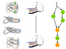
Discovery and Detection of Surgical Activity in Percutaneous VertebroplastiesIn this project, we aim at discovering automatically the workflow of percutaneous vertebroplasty. The medical framework is quite different from a parallel project , where we analyze laparoscopic surgeries. Contrary to cholecystectomies where much information is provided by the surgical tools and by the endoscopic video, in vertebroplasties and kyphoplasties, we believe that the body and hand movement of the surgeon give a key insight into the surgical activity. Surgical movements like hammering of the trocar into the vertebra or the stirring of cement compounds are indicative of the current workflow phase. The objectives of this project are to acquire the workflow related signals using accelerometers, processing the raw signals and detecting recurrent patterns in order to objectively identify the low-level and high-level workflow of the procedure. |
