Image-Based Glioma Modelling
This project consists of two main parts.
- PART 1: Development of Computational Models
- PART 2: Model Personalizations
- This includes simulations of glioma behavior in patient individual brain anatomy, estimation of model parameters and corresponding uncertainties.
Tumor Induced Brain Deformations

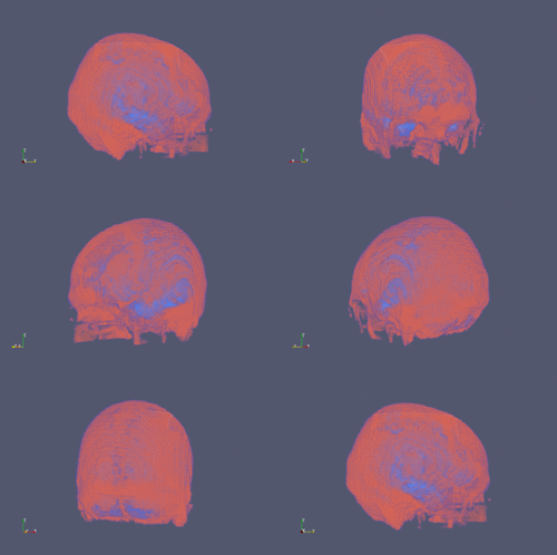
Reconstructed skull (low resolution data)
|
|
Increased intracranial pressure is the most critical symptom of brain cancer, leading to brain deformation, tissue and nerve compression. We present novel approach for modelling tumour progression together with corresponding brain deformations in patient individual anatomies obtained from medical images. We use the diffuse domain approach to implicitly capture complex brain anatomy through an auxiliary function. The governing equations are then appropriately modified and extended to a larger, regular domain. This approach helps to reduce errors caused by anatomy missegmentation and allows the use of efficient numerical methods that would not be applicable to the original problem formulation.
|
|
Patient Specific Glioma Modelling

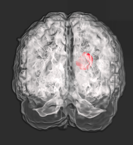
Simulated tumour in patient brain anatomy
|
|
Glioma is the most common type of brain tumors. It is characterised by infiltrative growth into the surrounding tissue, instead of forming solid tumours with well defined boundary. As a consequence only a part of the glioma beyond a certain concentration threshold is visible in MR images, while areas with lower tumour concentration appear normal on current imaging modalities. Such uncertainties in tumour shape and cell concentration complicate determination of optimal treatment plan.
We present a personalised, image based approach for inferring spatial distribution of glioma cells beyond limitations of medical images. The presented framework includes simulation of glioma growth and proliferation in patient individual brain anatomy reconstructed from medical images. Model parameters for each patient are inferred from multimodal images, including MRI scans and metabolic maps, using a Bayesian uncertainty quantification and parameter estimation framework. Uncertainties in model parameters are further propagated to the predictions of tumour shape and cell concentration. Efficiency of the inference is based on parallel Transitional Markov Chain Monte Carlo method. Clinical application of presented personalised approach lies in capability to provide complementary information to medical images.
|
|


