Workflow
- One-click segmentation of the aorta in a CTA (implementation by Oliver Kutter)
- Fiducial based calibration and projection matrix determination using the Direct Linear Transformation
- Distortion correction of the image intensifier
- Segmentation of the CT spots for the determination of their centroids in the three- and the two-dimensional space
- Determination of the internal and external orientation of the C-arm using the DLT
- Spatial position recovery of the intraoperative position of the Stent out of one x-ray image
- Interactive localization of the stent in the x-ray image
- Back projection of the stent - Definition of a ray of sight
- Visualization of the real stent position in a virtual CT volume
- Visual overlapping of a DRR (Digitaly Reconstructed Radiograph) of the CT volume and the particula x-ray image
- Effective update of the position of the Stent during the operation, regarding the right direction and depth in the Aorta
- GUI (implementation by Oliver Kutter)
Purpose
An aortic aneurysm is a local dilatation at the thoracic (15%) or abdominal (85%) part of the aorta, caused due to the thinning of the inner aortic wall. The endovascular stenting is a minimally invasive approach for the treatment of aortic aneurisms and it involves the exclusion of the aneurysm of the circulatory system with the placement of an implant, the stent. In the current clinical workflow, a CTA (Computed Tomography Angiography) scan of the region of interest is acquired some days before the operation. It helps the surgeon to exactly detect the aneurysm and the aortic bifurcations on the aorta (especially the renal arteries and the arteries on the arch for the blood supply of the head (brachiocephalic trunk, left common carotid, left subclavian), calculate its dimensions and select the appropriate implant, based on the length of the aneurysm and the diameter of the aorta at that place. Intraoperative, the stent is introduced from the femoral through the iliac arteries till the region of interest in the aorta. It is constructed with a self-attachment mechanism and, after it is unfolded, it replaces the inner wall of the aorta. A mobile fluoroscope, a C-arm, is used to support the procedure of the navigation towards the aneurysm and the positioning task of the implant. For highlighting the vessels, contrast agent is injected in the circulatory system during every x-ray image acquisition process. For the implantation of a single stent, ten to fifteen x-ray image series are required. However, the frequent use of the C-arm increases the radiation exposure in the operating room and the number of injections of contrast, which is very harmful for the patient. In this thesis a system for intraoperative three dimensional visualization of the patient’s anatomy, together with the actual stent position in the aorta and for navigation towards the aneurysm is proposed, with the objective of more accurate implantation and minimization of x-ray image acquisition.Requirements of the System
For the reasonable application of the system in the operating room, some preconditions have to be fulfilled- A preoperative CTA scan and a mobile fluoroscope have to be available. By means of these image acquisition systems the calibration, registration, and visualization methods can be realized.
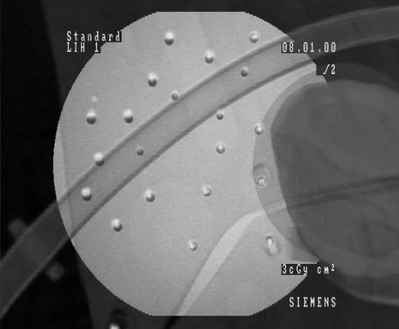
- CT spots have to be adhered on the patients skin at the region of interest, whereas not all of them must lie on the same plane (a couple of them are positioned e.g. on the back of the patient). They have to be visible in the CT scan and a minimum number of six of them in every x-ray image. Their position during the intervention has to be the same as during the CT scan.
- An efficient computer system for processing and visualization of large image data and real time applications has to be present in the operating room. The minimum requirements are a CPU of 2.0 MHz, main memory of 512 MB and graphic hardware of 128 MB of memory and a texture memory capable of three dimensional mapping.
Medical Image Segmentation
Implementing a fiducial based calibration and registration, the detection of the CT spots in the CT scan and in any particular x-ray image is necessary. For the segmentation, two separate approaches have been considered, due to the different acquisition properties of the two image intensifiers (CT and C-arm). To summarize, thresholding , region growing and circular object detection are some of the applied algorithms. In the three dimensional space, the centroids are determined via weighted intensities and partial volume effects and in the two dimensional by calculating the gravity centers of detected circles. The 3D segmentation is performed preoperatively, whereas the 2D detection is realized online. The success of the detection algorithms lies in both spaces over 90%.Camera Calibration
For the calibration and the pose estimation of the camera four steps are required- An approach for correction of the distortion, present in x-ray images. However, distorted images can also be used, providing acceptable results by the back projection
- An algorithm, based on graph matching, determines corresponding CT spots in 2D and 3D space
- The DLT algorithm (Direct Linear Transformation) is executed online (after every new x-ray) and determines the particular projection matrix P of the camera
- The RQ decomposition algorithm separates the projection matrix in two marrices, containing the intrinsic and the extrinsic camera parameters
Stent Three Dimensional Position Recovery and Intraoperative Navigation
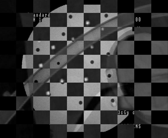
With position recovery we indicate the determination of a point (structure) in space, using one or more of its two dimensional projections (images). In this case we recover the position of the stent and visualize it in the volume rendered CT scan by using one intraoperative X-ray image. First, the segmented aorta is skeletonized and the one-voxel thick centerline is determined. Having computed the intrinsic and extrinsic parameters of the current projection, the particular image creation process can be defined. Thus, the position of the stent in the X-ray image can be back projected into the volume. The intersection of this ray of sight with the centerline of the aorta can represent the actual three dimensional stent position with a sub millimeter accuracy. For validation and visualization purposes, the visual overlay of the two available image modalities, the CT data and the X-ray image, has been implemented. After initializing the CT volume with the particular intrinsic and extrinsic camera parameters, a DRR is acquired and overlaid with the actual X-ray image. The overlapping of the CT spots in both images can be used to determine the accuracy of the system. Moreover, the aorta, visible in the CT (CTA) can be made visible in the X-ray image without the injection of additional contrast agent.
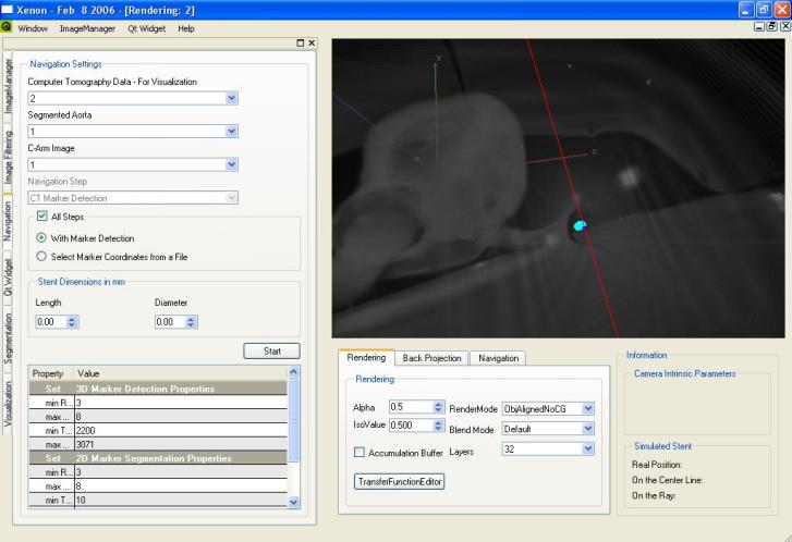
User interface of the application

Stent visualisation during the minimal invasive surgical treatment of a stenosis of the femoral artery.
Blue is the camera center (the source of the C-arm)


Visual overlay of the DRR and the X-ray image
| ProjectForm | |
|---|---|
| Title: | A Navigation Tool for the Endovascualar Treatment of Aortic Aneurysms - Computer Aided Implantation of a Stent Graft |
| Abstract: | More and more aneurysms of the thoracic and abdominal aorta are treated minimally-invasive by implanting a stent graft inside the aorta. However, without opening the patient the clinicians rely on imaging techniques, e.g. computed tomography, magnetic resonance imaging and X-ray, in order to visualize the region of interest and for intraoperative navigation. Unfortunately not all imaging modalities are available during operation, some are only available preoperatively. In 2003 the STENT project explored the prospects of using computer aided imaging techniques for preoperative planning and intraoperative navigation. Within the project a first prototype application was developed, but due to time constraints never made it to the operating room. This project is the continuation of the 2003's STENT project and its goal is to provide the clinicians with a preoperative planning tool and an intra-operative navigation and visualization tool. Preoperativly acquired computed tomography images can be used for segmentation, visualization and metric measurement of the aorta and aneurysm. Intra operatively taken X-ray images are aligned with the CT data, thus aiding navigation by supplying a three-dimensional visualization of an anatomical detailed model, metrics and current as well as planned locations of the stent graft. |
| Student: | Konstantinos Filippatos |
| Director: | Nassir Navab |
| Supervisor: | Marco Feuerstein |
| Type: | Diploma Thesis |
| Status: | finished |
| Start: | 2005/06/15 |
| Finish: | 2006/02/15 |
Edit |
Attach |
Refresh |
Diffs |
More |
Revision r1.31 - 27 Feb 2007 - 02:18 - KonstantinosFilippatos