Multiple Myeloma Staging using PET and CT images
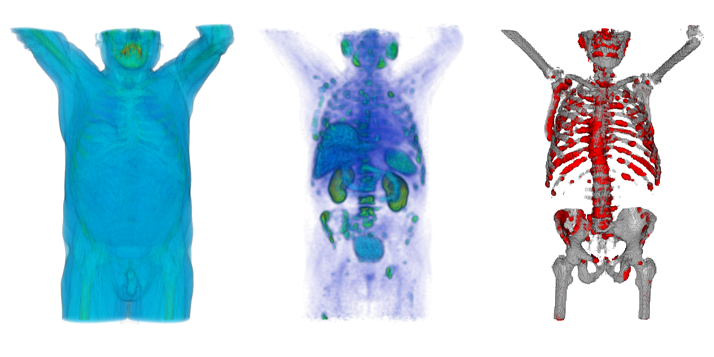
 Figure 1: Images of a prostata cancer patient with bone lesions. Left: CT, middle: PSMA-PET, right: bone lesions (red).
|
Project Description
Multiple Myeloma is a plasma cell cancer with affects 6.1 persons out of 100 000 every year. In further stages, the abnormal plasma cells accumulate in the bone marrow, which leads to bone lesions.The goal of this project is to use data from a Multiple Myeloma study to develop tools for fully automatic staging of the disease, and to model the disease progression using a large cohort of patients.
Tasks
- Incorporate information from PET and CT in order to automatically estimate bone tumor load
- Model the disease progression
- Validation of the model on clinical data
|
|
Requirements
- Good Programming Skills (Matlab or C++)
- Ability to work independently
- Experience with medical images processing is not required
Contact
If you are interested in the project or if you have any questions please contact
Materials & References
- Multiple Myeloma: some facts
- Staging using Blood values here
- Imaging in Multiple Myeloma here
- PET imaging: Positron emission tomography: basic sciences. D.L. Bailey, D.W. Townsend, P.E. Valk, and M.N. Maisey. Springer, 2004
 Figure 1: Images of a prostata cancer patient with bone lesions. Left: CT, middle: PSMA-PET, right: bone lesions (red).