Diploma thesis

|
Thesis by:
Advisor: Nassir Navab
Supervision by: Tassilo Klein, Ahmad Ahmadi
Due date: open
Abstract
Planning of a trajectory in neurosurgery (e.g. for biopsy or Deep Brain Stimulation, DBS) is a delicate task. Typically, a path is defined by analyzing a MRI image of the patient and setting an entry and a target point (in the case of a linear trajectory). The resulting linear path typically passes critical areas such as blood vessels or functional areas in very close proximity (e.g. <5mm). During surgery, after opening the skull and the protective dura mater of the brain, a pressure change and loss of cerebro-spinal fluid (CSF) can lead to a tissue shift of the brain – the so-called Brain Shift. Although this shift can be very small, e.g. 2mm in the deep brain areas, very little changes can deteriorate the reliability of the pre-operative plan significantly. In this thesis, we investigate how to use ultrasound for assessment and compensation of brain shift.Objectives
- A realistic model for brain shift should be developed based on a cryo-gel material (available), including thin tubes for the simulation of blood vessels. The cryo-gel allows ultrasound and MRI compatibility of the phantom (i.e. multi-modality).
- The brain phantom should be MRI scanned twice: in normal state and after a deformation was induced.
- An accuracy analysis for US-US registration compared to MRI-MRI registration has to be performed to asses feasibility of temporal brain shift detection (before and after opening of dura) intra-operatively using purely ultrasound.
- The possibility to deform pre-operative MRI based on intra-operative US should be investigated. For this, a MRI-US registration has to be implemented. Registration can be done 2D-3D or 3D-3D if 3D ultrasound is available. This aspect of the thesis is research-oriented.
- The quality of registration with burr-hole and transcranial ultrasound should be compared. Burr-hole ultrasound is simulated by leaving away the bone-mimicking tissue. Transcranial ultrasound is simulated by using several layers of bone-mimicking material (to be determined. e.g. PVC-U) with various thickness.
Literature
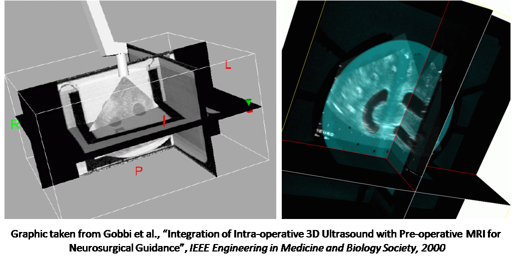
- Gobbi, D.; Comeau, R.; Lee, B. & Peters, T., Integration of intra-operative 3D ultrasound with pre-operative MRI for neurosurgical guidance, Engineering in Medicine and Biology Society, 2000. Proceedings of the 22nd Annual International Conference of the IEEE, 2000, 3, 1738-1740 vol.3
- Reinertsen, I. & Collins, D., A realistic phantom for brain-shift simulations, Medical Physics, 2006
- Reinertsen, I.; Descoteaux, M.; Siddiqi, K. & Collins, D., Validation of vessel-based registration for correction of brain shift, Medical Image Analysis, 2007, 11, 374-388
| Students.ProjectForm | |
|---|---|
| Title: | Ultrasound Registration for Brain Shift Analysis Using a Cryo-Gel Phantom |
| Abstract: | Planning of a trajectory in neurosurgery (e.g. for biopsy or Deep Brain Stimulation, DBS) is a delicate task. Typically, a path is defined by analyzing a MRI image of the patient and setting an entry and a target point (in the case of a linear trajectory). The resulting linear path typically passes critical areas such as blood vessels or functional areas in very close proximity (e.g. <5mm). During surgery, after opening the skull and the protective dura mater of the brain, a pressure change and loss of cerebro-spinal fluid (CSF) can lead to a tissue shift of the brain – the so-called Brain Shift. Although this shift can be very small, e.g. 2mm in the deep brain areas, very little changes can deteriorate the reliability of the pre-operative plan significantly. In this thesis, we investigate how to use ultrasound for assessment and compensation of brain shift. </font> |
| Student: | |
| Director: | Nassir Navab |
| Supervisor: | Tassilo Klein, Ahmad Ahmadi |
| Type: | DA/MA/BA |
| Area: | Registration / Visualization, Medical Imaging |
| Status: | finished |
| Start: | |
| Finish: | |
| Thesis (optional): | |
| Picture: | |