Master Thesis: Calculation of signal intensity-time curves from MR images of the beating heart
|
Thesis by: Radhika Tibrewal Advisors: Prof. Nassir Navab Supervision by: Michael Friebe as liasion to VISUS from CAMP and TUM-IAS Prof. Dr. Thomas Hackländer from HELIOS Klinikum, Wuppertal Dr. Abouzar Eslami from CAMP PDF of the thesis call |

|
Overview
Topic
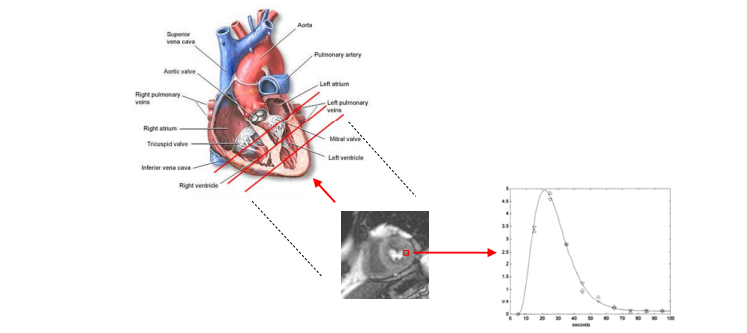
In order to assess whether a patient with a narrowing of a coronary artery has an increased risk for a heart at-tack, one is interested in quantitative parameters of the blood supply to the heart muscle. Currently those parameters could be judged only semi quantitative by visual analysis of a sequence of MRI images: For that approximately 300 consecutive dynamic images are acquired by a MRI (magnetic resonance imaging) scanner. During the acquisition an intravasal CM (contrast medium) is injected and the distribution of the signal intensity increase caused by the CM is visually observed by a radiologist. The higher the increase of the observed signal intensity, the lower the risk of the patient is to suffer a hard attack in the future. To derive reliable statistical data from these measurements, the increase of the signal intensity must be measured quantitatively in each pixel of the image. That means that the signal intensity-time curves must be derived automatically for each pixel for a time period of approximately 1 minute. Due to the breathing and small movements of the patients during the acquisition period, the measured images could not be evaluated directly. The main objective of this work is to develop a mathematical method to “freeze” the movement of the heart. The second object should be to calculate the signal intensity-time curve in each pixel of the frozen sequence and visualize the result as a parameter image of the signal intensity peak.Further Details
- This is an exciting project that involves MRI imaging, real clinical and scientific questions, and a fair amount of Computer Science.
- The project involves working within the R&D infrastructure of one of the most successful imaging companies.
- The project language is JAVA.
- The expectation is that the student spends at least some time in Bochum and Wuppertal -- minimum 3 months up to almost the entire time.
- An outline could be: 1 or 2 weeks initially to get to know the people and technical infrastructure, then maybe a month or two in Munich, then one month here, then a month in Munich and finally one or two month in Bochum again.
- The company would provide an apartment free of charge plus reasonable travel expenses and a € 400 allowance per month
- The clinical partner will provide the MRI data and will supervise the student in all medical questions.
| Students.ProjectForm | |
|---|---|
| Title: | Calculation of signal intensity-time curves from MR images of the beating heart |
| Abstract: | In order to assess whether a patient with a narrowing of a coronary artery has an increased risk for a heart at-tack, one is interested in quantitative parameters of the blood supply to the heart muscle. Currently those parameters could be judged only semi quantitative by visual analysis of a sequence of MRI im-ages: For that approximately 300 consecutive dynamic images are acquired by a MRI (magnetic resonance imaging) scanner. During the acquisition an intravasal CM (contrast medium) is injected and the distribution of the signal intensity increase caused by the CM is visually observed by a radiologist. The higher the increase of the observed signal intensity, the lower the risk of the patient is to suffer a hard attack in the future. To derive reliable statistical data from these measurements, the increase of the signal intensity must be measured quantitatively in each pixel of the image. That means that the signal intensity-time curves must be derived automatically for each pixel for a time period of approximately 1 minute. Due to the breathing and small movements of the patients during the acquisition period, the measured images could not be evaluated directly. The main objective of this work is to develop a mathematical method to “freeze” the movement of the heart. The second object should be to calculate the signal intensity-time curve in each pixel of the frozen sequence and visualize the result as a parameter image of the signal intensity peak. |
| Student: | Radhika Tibrewal |
| Director: | Prof. Nassir Navab |
| Supervisor: | Dr. Abouzar Eslami from CAMP affil. Prof. Dr. Michael Friebe as liasion from CAMP and TUM-IAS |
| Type: | Master Thesis |
| Area: | Medical Imaging |
| Status: | finished |
| Start: | |
| Finish: | |
| Thesis (optional): | |
| Picture: | |