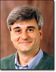
Visit of Prof. Benoit Dawant
Title
Visit of Prof. Benoit Dawant
Visitors
- Benoit Dawant - Professor of Electrical Engineering, Biomedical Engineering and Computer Science and head of the Medical Image Processing Laboratory at Vanderbilt University
Time and Location
Talk: during CAMP lecture, Tue. 25. Nov. 2008, CAMP seminar room, 13:00-14:30 o'clock
Links
Website of Prof. Benoit Dawant at Vanderbilt University
Website of the Medical Image Processing Laboratory at Vanderbilt University
Abstract
Prof. Dawant is visiting us in the course of the ROBOCAST project, as an external expert on computer-aided placement of Deep Brain Stimulation (DBS) electrode probes.
Here is an abstract of his talk:
Deep brain stimulation (DBS) is now considered by many neurologists and neurosurgeons as medically indicated in many patients with Parkinson’s disease, essential tremor, or dystonia in which medications are ineffective or no longer effective. In addition to appropriate patient selection, the surgical process of implantation must target and place the DBS lead so that at least one contact is within 1-2 mm of the intended target. Accurate targeting and placement is difficult because neurosurgeons must account for anatomical variability among patients, which can be significant. It is difficult even for experienced neurosurgeons to account for such three-dimensional changes by visual inspection of two-dimensional images, even with present day high resolution MR images. Following implantation, effective use of the device requires an experienced clinician to assess the location of each of the contacts with respect to the desired stimulation response as well as side effects so as to program the device with a setting that is therapeutically effective and hopefully energetically efficient. Device programming consists in selecting parameters (voltage, frequency, etc.), which create an electric field around the electrode. This field must be such that it affects regions of the brain responsible for the symptoms while not affecting regions, which may generate undesirable side effects.
In this talk, the status of an ongoing project at Vanderbilt University aiming at facilitating the entire procedure will be presented. Components of the system include a central repository in which data for over 200 cases performed over the last five years have been stored, automatic algorithms to create anatomic and electrophysiological atlases, automatic creation of pre-operative plans, automatic segmentation of anatomical structures, and visualization methods to assist in the placement and programming of the electrodes. The system has been integrated in the clinical flow and is currently under evaluation by an interdisciplinary team of engineers, neurosurgeons, electrophysiologists, and neurologists.

