ProjectStenosisClassification

Chair for Computer Aided Medical Procedures & Augmented Reality
Lehrstuhl für Informatikanwendungen in der Medizin & Augmented Reality

THIS WEBPAGE IS DEPRECATED - please visit our new website

Stenosis Classification Using Joint MR Intensities

Student: Benjamin Zaminer
Advisor: Nassir Navab, Holger Poppert, M.D. (Klinikum rechts der Isar, Neurology Dept.)
Supervision by: Andreas Keil

Abstract

Stroke is the third leading cause of death in Germany. It is a neurology injury, whereby the oxygensupply to parts of the brain gets cut off. About 80% of these strokes are due to ischemia, i.e. an occlusion of a bloodvessel leading to an interrupted blood flow.
MRWeightings.png
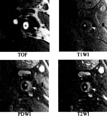
Stenosis inside the carotid artery imaged using four different MR weightings

Special setting in this project is the arteria carotis. Plaque is most likely to develop at the branching of the arteria carotis communis into the arteria carotis interna (leading to the brain) and the arteria carotis externa. This can lead to an abnormal narrowing, called a stenosis. According to the American Heart Association these plaques can be divided into different types, based on their consistency and structure.

Until now the decision about a surgery was only based on the degree of the stenosis and not on the type of plaque causing it. This is a faulty approach since there is a plaquetype (Type IV) which constitutes a relevant clinical danger, although it does not necessary come along with a stenosis. Unlike most other image modalities MR images do not only give information about the degree of the stenosis, but also about the consstency of the plaque. Using different weighted MR images it is possible to correctly classify plaque into the types defined by the AHA.

The main goal of this project is to create a classificationtool based on T1, T2, Proton Density and 'Time of flight' weighted images. To achieve this goal the arteria carotis and the plaqeue have to be segmented from the images. Furthermore various features of the plaque have to be extracted in order to get information needed for the classification.

Progress

Literature

  • [Nighoghossian2005] Norbert Nighoghossian, Laurent Derex, and Philippe Douek: The Vulnerable Carotid Artery Plaque, Stroke, 36:2764-2772, 2005, doi:10.1161/01.STR.0000190895.51934.43
  • [Clarke2006] Sharon E. Clarke, Vadim Beletsky, Robert R. Hammond, Robert A. Hegele, and Brian K. Rutt: Validation of Automatically Classified Magnetic Resonance Images for Carotid Plaque Compositional Analysis, Stroke, 37:93-97, 2006, doi:10.1161/01.STR.0000196985.38701.0c

Students.ProjectForm
Title: Stenosis Classification Using Joint MR Intensities
Abstract:  
Student: Benjamin Zaminer
Director: Prof. Dr. Nassir Navab
Supervisor: Andreas Keil
Type: Project
Area:  
Status: finished
Start: 15/05/2006
Finish: 01/02/2008


Edit | Attach | Refresh | Diffs | More | Revision r1.24 - 27 Feb 2009 - 21:39 - AndreasKeil