Stent Graft Detection in 2D Xray Images
Advisor: Nassir NavabSupervision by: Stefanie Demirci, Ali Bigdelou

Overview
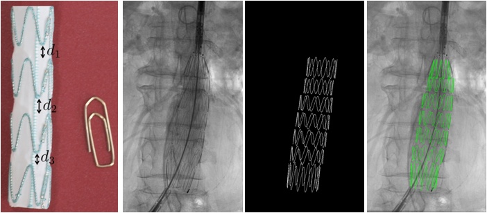
In the current clinical workflow of endovascular abdominal aortic repairs (EVAR) a stent graft is inserted via an introducer system through one femoral artery into the aneurysmatic aorta under 2D angiographic imaging. Due to the missing depth information in the X-ray visualization, it is highly difficult in particular for junior physicians to place the stent graft in the preoperatively defined position within the aorta. Therefore, methods for accurate stent graft recognition or segmentation in fluoroscopy images are highly required. In this project, you are supposed to adapt an existing MATLAB algorithm to new requirements. This new implementation should then be correctly validated in an animal study. It is your task to help us with the plan of the experimental setup in cooperation with our colleagues at the university hospital lab NARVIS as well as with our partner physicians. You should have very good knowledge of MATLAB programming and should be able to understand very well commented code. It would be beneficial for the experiment planning if you are interested in medical applications and can interact with people from different disciplines smoothly. If you are interested please send a brief CV as well as a sample of MATLAB code that you have already implemented, to demirci@in.tum.de.Tasks
- Adaptation of existing MATLAB algorithms to new requirements.
- Validation of method on synthetic test data and real patient data.
- If interested: Organization of animal study at our hospital lab NARVIS in collaboration with Dr. Ghotbi (Kreisklinik München-Pasing) and our partner physicians from Klinikum Innenstadt.
Requirements
- A good knowledge of MATLAB is mandatory.
- Interest in medical applications and multidisciplinary work is required.
| ProjectForm | |
|---|---|
| Title: | Stent Graft Detection in 2D Xray Images |
| Abstract: | In the current clinical workflow of endovascular abdominal aortic repairs (EVAR) a stent graft is inserted via an introducer system through one femoral artery into the aneurysmatic aorta under 2D angiographic imaging. Due to the missing depth information in the X-ray visualization, it is highly difficult in particular for junior physicians to place the stent graft in the preoperatively defined position within the aorta. Therefore, methods for accurate stent graft recognition or segmentation in fluoroscopy images are highly required. |
| Student: | Radhika Tibrewal |
| Director: | Prof. Dr. Nassir Navab |
| Supervisor: | Stefanie Demirci, Ali Bigdelou |
| Type: | SEP |
| Area: | Registration / Visualization, Segmentation |
| Status: | finished |
| Start: | |
| Finish: | |
| Thesis (optional): | |
| Picture: | |