Diploma thesis

|
Thesis by:
Advisor: Nassir Navab
Supervision by: Tassilo Klein, Ahmad Ahmadi
Due date:
Abstract
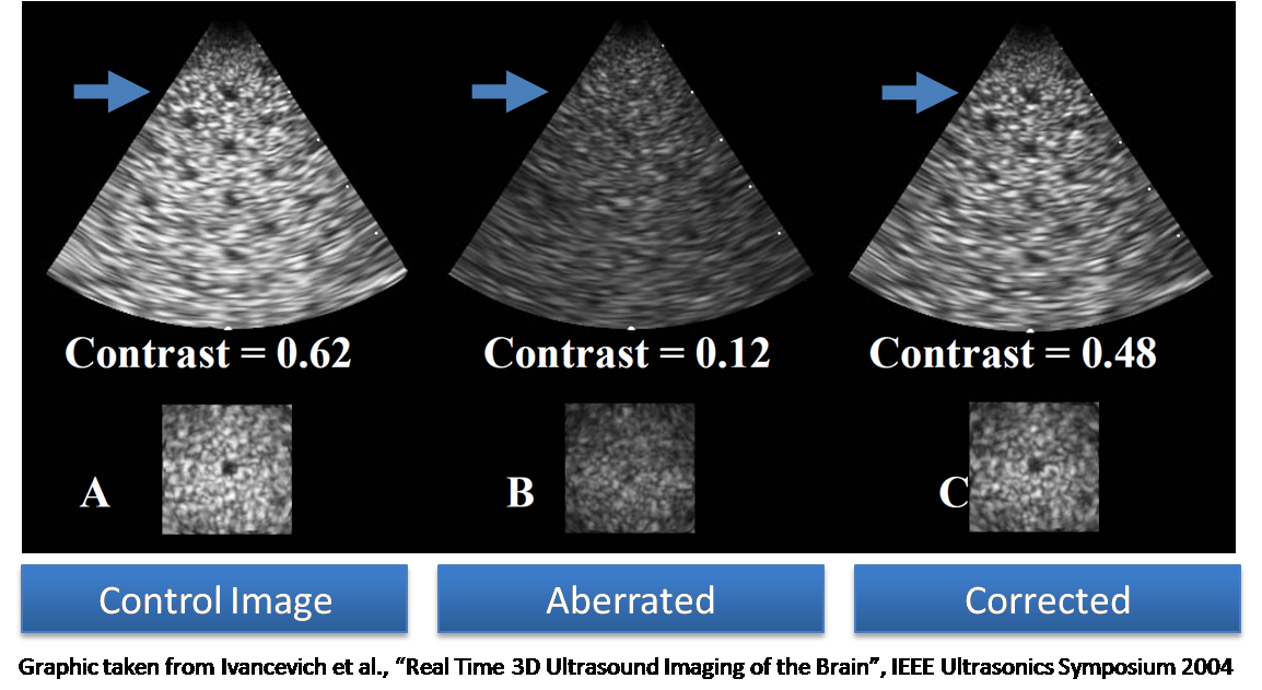
Ultrasound is a non-invasive imaging technique that can be easily and cheaply integrated into the OR environment. We plan to use ultrasound for neurosurgical interventions. Transcranial ultrasound means imaging the brain through the skull of patient (i.e. through the temporal lobe, or temples, in German “Schläfe”) of the patient. This technique is especially appealing because it is entirely non-invasive and even does not require sterilization. However, scanning through human bone modifies the ultrasound waves by changing their phase, hence the name “phase aberration”. Recent literature suggests that this “phase aberration” can be corrected. For that, we track the ultrasound optically and derive the skull thickness at the scanned position from a previous CT or MRI image.Objectives
- Phase aberration correction based on the thickness of penetrated bone tissue has to be implemented. This aspect
- A validation phantom has to be built: e.g. several nylon strings in water bath, with defined distances. Several layers of bone-mimicking material (to be determined. e.g. PVC-U) with various thickness will serve to simulate the skull bone.
- MRI, CT and according ultrasound data of one or several humans will be acquired before or parallel to the thesis for experimentation with real-life data (together with our medical partners at Klinikum Grosshadern).
- The evaluation: scanning the phantom (with known geometry) through "bone windows" with increasing thicknesses. Several layers of bone-mimicking material (to be determined. e.g. PVC-U, Silicon) with various thickness will be created for this step.
- Evaluate FRE accuracy with 2MHz, 3MHz and various bone window thicknesses, WITHOUT and then WITH Phase aberration correction --> does the accuracy of localization improve?
Expected Results:
- Ideally, the quality of transcranial ultrasound images should improve, allowing a better localization and imaging of brain tissue as well as vasculature.
Literature
- Hynynen, K. & Sun, J., Trans-skull ultrasound therapy: the feasibility of using image-derived skull thickness information to correct the phase distortion, Ultrasonics, Ferroelectrics and Frequency Control, IEEE Transactions on, 1999, 46, 752-755
- Light, E.; Smith, S.; Ivancevich, N. & Dahl, J., 2B-2 Phase Aberration Correction on a 3D Ultrasound Scanner Using RF Speckle from Moving Targets, Ultrasonics Symposium, 2006
- Ivancevich, N.; Pinton, G.; Smith, S. & Nicoletto, H., 7A-5 Real-Time 3D Contrast-Enhanced Transcranial Ultrasound, Ultrasonics Symposium, 2007
| Students.ProjectForm | |
|---|---|
| Title: | Phase Aberration Compensation for Transcranial Ultrasound |
| Abstract: | Ultrasound is a non-invasive imaging technique that can be easily and cheaply integrated into the OR environment. We plan to use ultrasound for neurosurgical interventions. Transcranial ultrasound means imaging the brain through the skull of patient (i.e. through the temporal lobe, or temples, in German “Schläfe”) of the patient. This technique is especially appealing because it is entirely non-invasive and even does not require sterilization. However, scanning through human bone modifies the ultrasound waves by changing their phase, hence the name “phase aberration”. Recent literature suggests that this “phase aberration” can be corrected. For that, we track the ultrasound optically and derive the skull thickness at the scanned position from a previous CT or MRI image. |
| Student: | |
| Director: | Nassir Navab |
| Supervisor: | Tassilo Klein, Ahmad Ahmadi |
| Type: | DA/MA/BA |
| Area: | Medical Imaging |
| Status: | draft |
| Start: | |
| Finish: | |