Computer assisted optical biopsy for colorectal polyps.
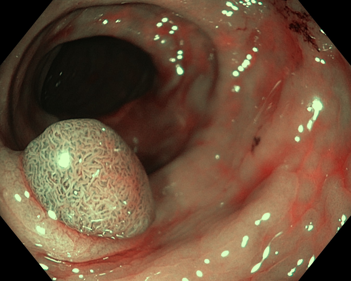
- Adenom

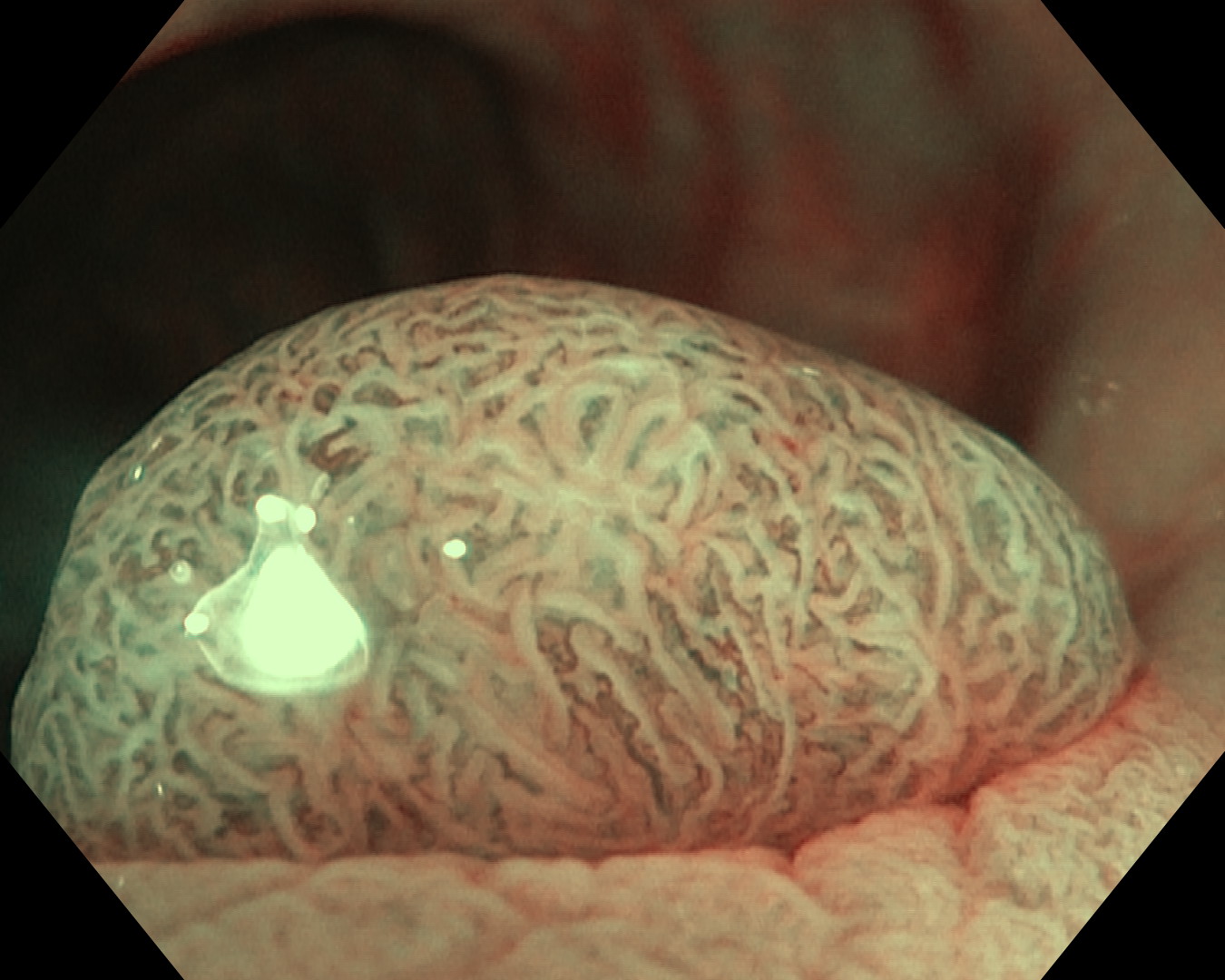
- Adenom

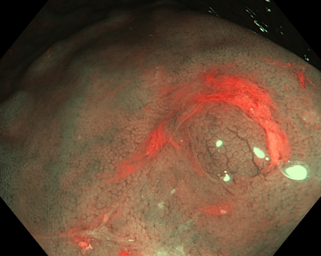
- Non-adenomatous Polyp

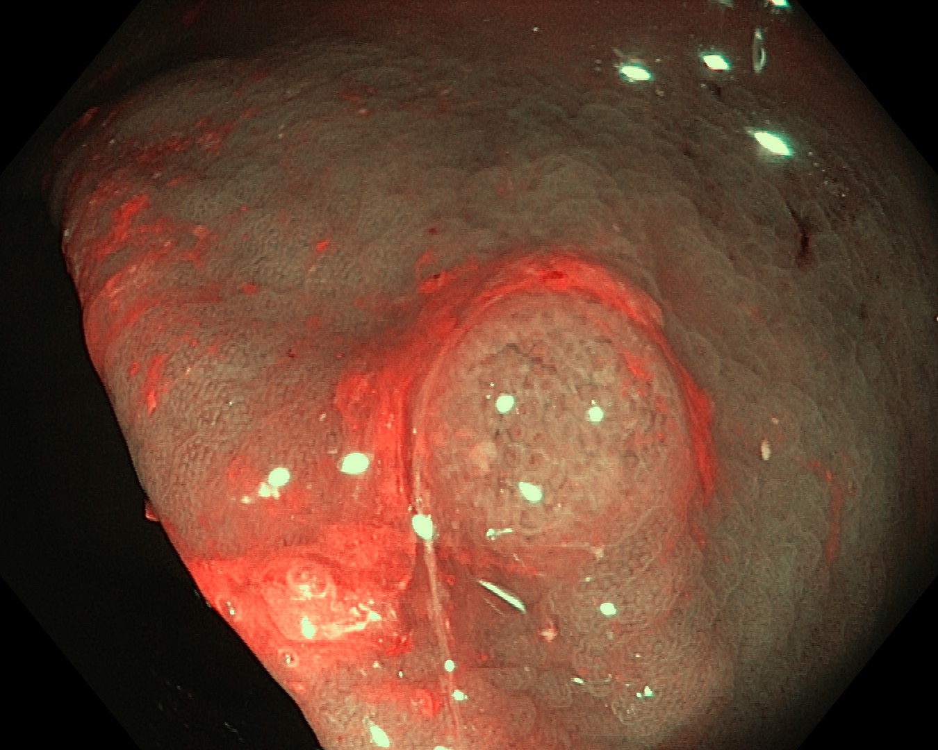
- Non-adenomatous Polyp

Figure 1: Examples of adenomas and non-adenomatous polyps.
|
Overall Project Description
This is a project in collaboration with the Klinikum Rechts der Isar. The difficulty of the project will be adapted to an IDP, Bachelor or Master Thesis.
The problem to address is the classification of images of colorectal polyps in two classes: Adenoma or non-adenomatous polyp (See figure).
From a series of optical and NBI(Narrow Band Imaging) images acquired in the clinic and labelled with the help of a Pathologist, the aim of the project is to identify suitable image features and learn a classifier able to automatically predict the class of a polyp given a new image. Challenges include the similarity of the two classes in some cases, poor imaging conditions (e.g. specular reflections) as well as a limited amount of labelled images.
To solve the problem we propose to study different feature extraction techniques and rely on machine learning methods (e.g. Random Forests) for learning the classifier.
Clinical Motivation
Adenomas are polyps of the colorectum that have the potential to develop into colon cancer [1]. However, some adenomas never become malignant tumors and if they do, progression from adenoma into cancer usually takes a long time. As a result, screening colonoscopy programs were established in order to detect and resect adenomas at an early stage [2]. After resection, polyps should be sent to pathology in order to make a histological diagnosis. Not every colorectal polyp has adenomatous histology. Approximately 40-50% of all polyps contain other benign histology (e.g. hyperplastic or inflammatory polyps). These polyps do not bear the risk of colon cancer.
The implementation of screening programs has led to increasing numbers of colonoscopies in the last years [3]. This approach naturally implies higher amounts of detected polyps. The removal of these polyps and consultation of a pathologist in order to make a diagnosis is time consuming and expensive. An optical-based prediction of polyp histology (adenomatous versus non- adenomatous) would enable endoscopists to save money and to inform patients faster about examination results. The approach of predicting polyp histology on the basis of optical features is called the “optical biopsy” method. The prediction is made by the endoscopists during real-time colonoscopy. The aim of this strategy is to make an optical diagnosis which enables users to resect polyps without sending the specimen to pathology. Narro Band Imaging (NBI) is a light-filter device which can be switched on during colonoscopy. NBI is useful to better display vascular patterns of the colon mucosa. It has been shown that the use of NBI can facilitate optical classification of colorectal polyps [5]. A NBI- based classification schemes exists which can be used to assign polyps into specific polyp categories (adenomatous versus non- adenomatous) [6].
Prior to the implementation of the optical classification approach for routine use in endoscopy it is necessary to proof its feasibility and accuracy [7]. Otherwise the approach would entail the risk of wrong diagnoses which could lead to wrong recommendations on further diagnostic or therapeutic steps.
Until now, some clinical trials have shown good accuracy for the optical biopsy method [5]. However, there is growing evidence that optical biopsy does not yet meet demanded accuracy thresholds [8]. The aim of the project “Computer assisted optical biopsy in the colorectum” is to create a computer program that is able to distinguish between adenomatous and non-adenomatous polyps. Still images of colorectal polyps including NBI- pictures of polyps will be used for machine learning.
Tasks
The main technical tasks to achieve are:
- Study and implement suitable feature extraction techniques
- Learn classifiers able to predict the polyp class
- Quantitative Validation according to expert labelled image dataset
According to the advancement, we may include learning and prediction from incomplete data, e.g. with semi-supervised learning
|
|
Requirements
- Good programming skills in MATLAB
- Programming skills in C++ are an advantage.
- Basic knowledge of image processing are recommendable.
Contact
If you are interested in the project or if you have any questions please contact
Diana Mateus
References
- Vogelstein B, Fearon ER, Hamilton SR, Kern SE, Preisinger AC, Leppert M, et al. Genetic Alterations during Colorectal-Tumor Development. New England Journal of Medicine. 1988; 319:525-32.
- Brenner H, Altenhofen L, Stock C, Hoffmeister M. Prevention, early detection, and overdiagnosis of colorectal cancer within 10 years of screeningcolonoscopy in Germany. Clin Gastroenterol Hepatol. 2015; 13:717-23.
- Stock C, Haug U, Brenner H. Population-based prevalence estimates of history of colonoscopy or sigmoidoscopy: review and analysis of recent trends. Gastrointest Endosc. 2010; 71: 366-381
- Lopez-Ceron M, Sanabria E, Pellise M. Colonic polyps: is it useful to characterize them with advanced endoscopy? World J Gastroenterol. 2014; 20: 8449-57.
- ASGE Technology Committee, Abu Dayyeh BK, Thosani N, Konda V, Wallace MB, Rex DK, Chauhan SS, Hwang JH, Komanduri S, Manfredi M, Maple JT, Murad FM, Siddiqui UD, Banerjee S. ASGE Technology Committee systematic review and meta-analysis assessing the ASGE PIVI thresholds for adopting real-time endoscopic assessment of the histology of diminutive colorectal polyps. Gastrointest Endosc. 2015 Mar;81(3):502.e1-502.e16
- Hewett DG, Kaltenbach T, Sano Y, Tanaka S, Saunders BP, Ponchon T, Soetikno R, Rex DK. Validation of a simple classification system for endoscopic diagnosis of small colorectal polyps using narrow-band imaging. Gastroenterology. 2012; 143: 599-607
- Kamiński MF, Hassan C, Bisschops R, Pohl J, Pellisé M, Dekker E, Ignjatovic-Wilson A, Hoffman A, Longcroft-Wheaton G, Heresbach D, Dumonceau JM, East JE. Advanced imaging for detection and differentiation of colorectal neoplasia: European Society of Gastrointestinal Endoscopy (ESGE) Guideline. Endoscopy. 2014; 46:435-49.
- Kang HY, Kim YS, Kang SJ, Chung GE, Song JH, Yang SY, Lim SH, Kim D, Kim JS. Comparison of Narrow Band Imaging and Fujinon Intelligent Color Enhancement in Predicting Small Colorectal Polyp Histology. Dig Dis Sci. 2015 Apr 14. [Epub ahead of print]


