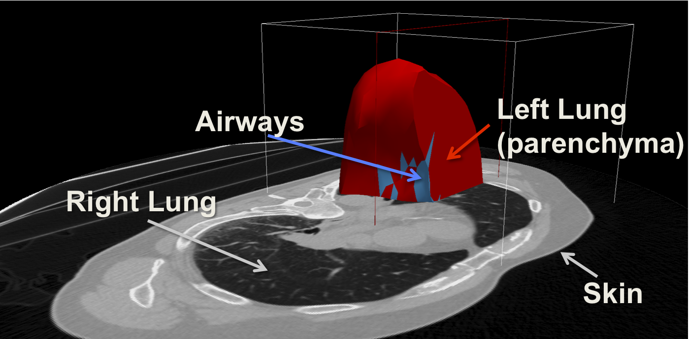
Computational Modeling of Respiratory Motion Based on 4D CT
Thesis by: Bernhard Fuerst Due date: June 15th, 2012Advisor: Prof. Nassir Navab
Supervision by: Mehmet Yigitsoy
Siemens Corporate Research Supervision by: Ali Kamen and Tommaso Mansi
Open Position
Type: HiWi at CAMP followed by an internship at Siemens Corporate Research in Princeton, New Jersey, USA (SCR)Start: HiWi: May; Internship: August 2012 (8 months)
Tasks: Developing the motion model framework (C++ and Matlab) and publication-oriented research.
Requirements: C++, Matlab, English, Teamwork
For more information or an application, send an e-mail with short introduction of yourself and CV to fuerst@in.tum.de (Subject: "MotionModelHiWi Your Name") until May 5th.
Abstract
Literature
- D.L. Pham, C. Xu, and J.L. Prince, “Current methods in medical image segmentation 1,” Annual review of biomedical engineering, vol. 2, no. 1, pp. 315–337, 2000.
- L. Grady, “Random walks for image segmentation,” Pattern Analysis and Machine Intel- ligence, IEEE Transactions on, vol. 28, no. 11, pp. 1768–1783, 2006.
- T. Kohlberger, M. Sofka, J. Zhang, N. Birkbeck, J. Wetzl, J. Kaftan, J. Declerck, and S. Zhou, “Automatic multi-organ segmentation using learning-based segmentation and level set optimization,” Medical Image Computing and Computer-Assisted Intervention– MICCAI 2011, pp. 338–345, 2011.
- N. Otsu, “A threshold selection method from gray-level histograms,” Automatica, vol. 11, pp. 285–296, 1975.
- H. Samet and M. Tamminen, “Efficient component labeling of images of arbitrary di- mension represented by linear bintrees,” Pattern Analysis and Machine Intelligence, IEEE Transactions on, vol. 10, no. 4, pp. 579–586, 1988.
- P. Risholm, J. Ross, G. Washko, and W. Wells, “Probabilistic elastography: Estimating lung elasticity,” in Information Processing in Medical Imaging. Springer, 2011, pp. 699–710.
- J.R. McClelland?, J.M. Blackall, S. Tarte, A.C. Chandler, S. Hughes, S. Ahmad, D.B. Lan- dau, and D.J. Hawkes, “A continuous 4d motion model from multiple respiratory cycles for use in lung radiotherapy,” Medical Physics, vol. 33, pp. 3348, 2006.
- P.F.Villard,M.Beuve,B.Shariat,V.Baudet,andF.Jaillet,“Simulationoflungbehaviour with finite elements: Influence of bio-mechanical parameters,” in Medical Information Visualisation-Biomedical Visualisation, 2005.(MediVis? 2005). Proceedings. Third International COnference on. IEEE, pp. 9–14.
- J. Eom, C. Shi, X. Xu, and S. De, “Modeling respiratory motion for cancer radiation therapy based on patient-specific 4dct data,” Medical Image Computing and Computer- Assisted Intervention–MICCAI 2009, pp. 348–355, 2009.
- M.P. Vijaykumar, A. Kumar, and S. Bhatia, “Latest trends, applications and innovations in motion estimation research,” International Journal of Scientific & Engineering Research, vol. 2, no. 7, 2011.
- S. Rit, J. Nijkamp, M. van Herk, and J.J. Sonke, “Comparative study of respiratory motion correction techniques in cone-beam computed tomography,” Radiotherapy and Oncology, 2011.
- AP King, C. Buerger, C. Tsoumpas, P. Marsden, and T. Schaeffter, “Thoracic respira- tory motion estimation from mri using a statistical model and a 2-d image navigator,” Medical Image Analysis, 2011.
- A. Al-Mayah, J. Moseley, and KK Brock, “Contact surface and material nonlinearity modeling of human lungs,” Physics in medicine and biology, vol. 53, pp. 305, 2008.
- K.K. Brock, A.M. Nichol, C. Ménard, J.L. Moseley, P.R. Warde, C.N. Catton, and D.A. Jaffray, “Accuracy and sensitivity of finite element model-based deformable registra- tion of the prostate,” Medical physics, vol. 35, pp. 4019, 2008.
- A. Al-Mayah, J. Moseley, M. Velec, and KK Brock, “Sliding characteristic and material compressibility of human lung: Parametric study and verification,” Medical physics, vol. 36, pp. 4625, 2009.
- A.S.Naini,ModelingLungTissueMotionsandDeformations:ApplicationsinTumorAblative Procedures, Ph.D. thesis, The University of Western Ontario, 2011.
- J. Ehrhardt, R. Werner, A. Schmidt-Richberg, B. Schulz, and H. Handels, “Generation of a mean motion model of the lung using 4d-ct image data,” in Eurographics Workshop on Visual Computing for Biomedicine. Citeseer, 2008, pp. 69–76.
- M.J.D. Powell, “Developments of newuoa for minimization without derivatives,” IMA journal of numerical analysis, vol. 28, no. 4, pp. 649, 2008.
- J.R. Shewchuk, “Tetrahedral mesh generation by delaunay refinement,” in Proceedings of the fourteenth annual symposium on Computational geometry. ACM, 1998, pp. 86–95.
| Students.ProjectForm | |
|---|---|
| Title: | Computational Modeling of Respiratory Motion Based on 4D CT |
| Abstract: | Respiratory lung motion has a serious impact on the quality of medical imaging, treatment planning and intervention, and radiotherapy. This motion not only reduces imaging quality, especially for positron emission tomography (PET), but also inhibits the determination of the exact position and shape of the target during radiotherapy. Based on prior knowledge of average tissue properties, patient-specific imaging (4D CT) and a surrogate signal, a computational motion model can be created. This enables researchers and developers to simulate and generate information about a respiratory phase not covered by the imaging procedure. Therefore, the internal deformation of the lung and its containing cancerous tissue can be computed and taken into account during further imaging acquisitions or radiotherapy. |
| Student: | Bernhard Fuerst |
| Director: | Prof. Nassir Navab |
| Supervisor: | Mehmet Yigitsoy |
| Type: | Master Thesis |
| Area: | Registration / Visualization |
| Status: | finished |
| Start: | 2011/11/15 |
| Finish: | 2011/06/15 |
| Thesis (optional): | Master Thesis |
| Picture: | |
 A biomechanical model - driven by estimated pressure values - allows the simulation of lung movements.