Available SEP / IPD in Medicine (max. 2 Students)

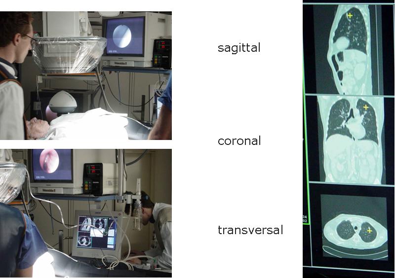
The figure shows a mock-up of the intervention with the electorimagnetic tracking device on the left and a mock-up for the user interface on the right. For further information on this SEP contact Joerg Traub.
Navigated Flexible Procedures (Bronchoscopy)
Image-directed bronchoscopy plays a major role in evaluating peripheral localized pulmonary lesions since they can not directly be visualized in the endoscope. Histopathological, cytological or microbiological assessment is essential in order to initiate adequate therapy. Different studies showed that the diagnostic yield of successful flexible bronchoscopy is between 18 to 62%. Normally, it is performed under X-ray imaging (a.k.a. fluoroscopy) that provides no depth perception, does not depict the anatomy of the bronchial tree and is even hardly able to discern the target. To improve the diagnostic yield, a precise navigation to the target region using a electromagnetic position and orientation (pose) measurement device (electromagnetic tracker) is used. An electromagnetic tracker generates an electromagnetic field and a coil sensor measures its position and orientation by the induction of current in the cois. An interface that provides the real-time pose of the sensor is provided. The navigation system that has to be implemented within this SEP has to visualize three orthogonal views of CT data and show a crosshair at the measured position of electromagnetic device in all three views. A point based registration method will be provided in order to register the coordinate system of the tracker with the patient's data. The implementation will be in C++ (VS Compiler) using OpenGL. An interface to access the real-time data of the electromagnetic tracking device will be provided, as well as a library to read the medical imaging data(DICOM) as recieved from the CT scanner.
The figure shows a mock-up of the intervention with the electorimagnetic tracking device on the left and a mock-up for the user interface on the right. For further information on this SEP contact Joerg Traub.
| ProjectForm | |
|---|---|
| Title: | Navigated Flexible Procedures (Bronchoscopy) |
| Abstract: | Bronchoscopy minimally invasive access to peripheral lung locations. The goal is to implement an application that visualizes within imaging data of computed tomography images the actual position of a electrimagnetic sensor. Therefore, a graphical user interface that visualizes three orthogonal sliceviews as well as a crosshair, that indicates the current position of the sensor. For tracking the Aurora system from Northern Digital will be used. |
| Student: | Julian Much |
| Director: | Nassir Navab & Prof. H. Feussner (MITI) |
| Supervisor: | Joerg Traub & Armin Schneider (MITI) |
| Type: | SEP |
| Status: | finished |
| Start: | 2006/02/01 |
| Finish: | 2006/06/01 |