Christoph Hennersperger
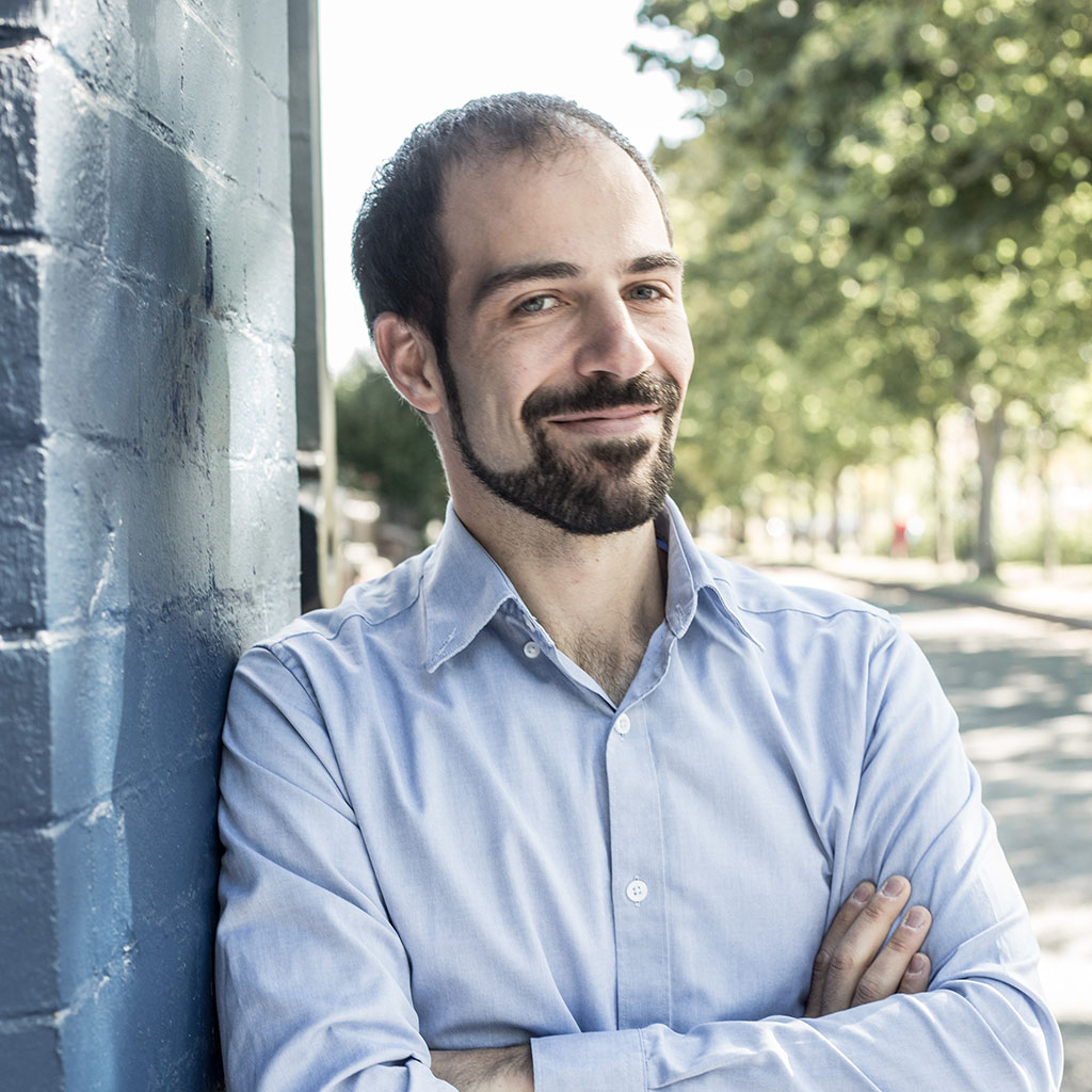 |
Dr. Christoph Hennersperger Co-Founder and CTO at OneProjects Affiliated senior research scientist and lecturer at CAMP Mail: christoph.hennersperger@tum.de OneProjects Munich, Germany & Dublin, Ireland |
Opportunities
We are continuously looking for motivated students for internships and student projects. Please check the OneProjects page for more information on the company and reach out to our team directly for more details and specific opportunities with us.Short Profile
I am passionate in medical device development, medical image computing, information technology, and improving healthcare. I enjoy working on challenging and exciting projects, integrating technical excellence and innovative thinking in truly collaborative teams. In 2017 I co-founded OneProjects, which operates at the intersection of hardware, software and data science to create the future vision for healthcare and how to successfully treat patients in a new era for data driven solutions.Curriculum Vitae:
- since 03/2017 - Founder and CTO at OneProjects, an Irish-German MedTech? startup in Cardiac imaging
- since 06/2016 - Director of the MedInnovate innovation fellowship
- 03/2017 - 02/2019 - Research fellow at Trinity College Dublin, Ireland
- 06/2016 - 06/2018 - Research manager of the Interdisciplinary Research Laboratory at Klinikum rechts der Isar, and lead of EU Horizon2020 Project EDEN2020
- 08/2015 - 05/2016 - Fellow at BioInnovate Ireland, a innovation program in MedTech affiliated to Stanford BioDesign
- 08/2011 - 08/2015 - Research scientist at CAMP, Dr. rer. nat. obtained 05/2015
- 08/2011 - 11/2013 - Researcher for 3D freehand Ultrasound at Curefab GmbH, Munich
- 10/2006 - 11/2011 - Studies in electrical enginering at TUM (Informationstechnik)
- 09/1998 - 07/2006 - Abitur
Supervised theses
- Enhanced 3D-Ultrasound Visualization via Structure-Geometrical Ray Casting(MSc, finished)
- 3-D Segmentation of the Head and Neck Lymph Nodes Using the Level Set Methods in Ultrasound (MSc, finished)
- Deformable Vessel Registration for 3D Freehand Doppler Ultrasound (BSc, finished)
- Deformation Estimation and Correction by Biomechanical Modeling (MSc, finished)
- Automatic Extraction of Useful Structures for Ultrasound-Guided Femoral Nerve Blocks (MSc, finished)
- Segmentation of Musculus Puborectalis in 4D OB/GYN ultrasound data (BSc, finished)
- Autonomous Robotic Ultrasound for Screening Applications (MSc, finished)
- Full color surgical 3D printed tractography (MSc, finished)
- Multi-modal Deformable Registration in the Context of Neurosurgical Brain Shift (MSc, finished)
- Seamless stitching of 4D optoacoustic data (MSc, finished)
- Sparse Data Reconstruction and its Application in Spherical Computational Sonography (MSc, ongoing)
- Optimal planning and data acquisition for robotic ultrasound-guided spinal needle injection (MSc, finished)
- Sensor System Development for Measuring and/or Characterizing Surgical Drain Fluid Output (Msc, finished)
- Implementation of Resting State fMRI Functional Connectivity Analysis for the Mapping of Functional Brain Networks in Neurosurgery Treatment Planning (MSc, finished)
- Automated Camera System for Laparoscopic Surgeries Supported by Surgical Data Science (MSc, finished)
- Restoring the sensation in an upper limb with a smart Orthesis (MSc, finished)
- Ultrasound-Based Tissue Characteriziation for Ablation Procedures (Msc, finished)
IDP
- Tensor-Graph based segmentation of ultrasound images (finished)
- 3D velocity field reconstruction from ultrasound doppler data (finished)
- Optimization of a quadratic energy minimization framework for arbitrarily sampled ray data (finished)
- Implementation of a force-impedance controller for ROS and KUKA LBR 4+ (finished)
Teaching
- WS2020/21: Computer Aided Medical Procedures I - Ultrasound Imaging
- WS2019/20: MedInnovate: from unmet clinical needs to solution concepts
- WS2019/20: Computer Aided Medical Procedures I - Ultrasound Imaging
- WS2018/19: MedInnovate: from unmet clinical needs to solution concepts
- SS2018: MedInnovate: from unmet clinical needs to solution concepts
- WS2017/18: MedInnovate: from unmet clinical needs to solution concepts
- SS2017: BioInnovation: from unmet clinical needs to solution concepts
- SS2017: Brain Tumor Treatment - Ethical Standards, Medical challenges, Technological Future
- WS2016/17: BioInnovation: from unmet clinical needs to solution concepts
- WS2016/17: Image-based tool tracking for computer aided biopsy procedures
- WS2015/16: BioInnovation: from unmet clinical needs to solution concepts
- WS2015/16: Crowdsourcing and Gamification with Applications to Computer Vision
- SS2015: From an Unmet Clinical Need to a Promising Concept - FUN2Concept
- SS2015: Innovations in Robotic Imaging
- WS2014/15: Project Management and Software Development for Medical Applications
- WS2014/15: Innovations in Computer Aided Surgery
- SS2014: Innovations in Medical Robotics
- WS2009/10: Praktikum C++
Publications
| 2019 | |
| J. Rackerseder, M. Baust, R. Göbl, N. Navab, C. Hennersperger
Landmark-Free Initialization of Multi-Modal Image Registration Bildverarbeitung fuer die Medizin (BVM), Luebeck, March 17-19, 2019. (bib) |
|
| M. Esposito, C. Hennersperger, R. Göbl, L. Demaret, M. Storath, N. Navab, M. Baust, A. Weinmann
Total Variation Regularization of Pose Signals with an Application to 3D Freehand Ultrasound IEEE Transactions on Medical Imaging (TMI), Volume 38, Issue 10, pp 2245-2258, October 2019. (bib) |
|
| 2018 | |
| J. Rackerseder, M. Baust, R. Göbl, N. Navab, C. Hennersperger
Initialize globally before acting locally: Enabling Landmark-free 3D US to MRI Registration 21st International Conference on Medical Image Computing and Computer Assisted Interventions (MICCAI), Granada, Spain, September 2018 (bib) |
|
| B. Busam, P. Ruhkamp, S. Virga, B. Lentes, J. Rackerseder, N. Navab, C. Hennersperger
Markerless Inside-Out Tracking for 3D Ultrasound Compounding International Conference on Medical Image Computing and Computer Assisted Interventions (MICCAI), Point-of-Care Ultrasound, Granada, Spain, September 2018 [oral]. (bib) |
|
| R. Göbl, N. Navab, C. Hennersperger
SUPRA: open-source software-defined ultrasound processing for real-time applications International Journal of Computer Assisted Radiology and Surgery / 9th International Conference on Information Processing in Computer-Assisted Interventions (IPCAI), Berlin, Germany, June 2018. The original publication is available online at link.springer.com (bib) |
|
| S. Virga, R. Göbl, M. Baust, N. Navab, C. Hennersperger
Use the Force: Deformation Correction in Robotic 3D Ultrasound International Journal of Computer Assisted Radiology and Surgery / 9th International Conference on Information Processing in Computer-Assisted Interventions (IPCAI), Berlin, Germany, June 2018. The original publication is available online at springer.com (bib) |
|
| S. Nitkunanantharajah, C. Hennersperger, X.L. Dean-Ben, D. Razansky, N. Navab
Trackerless panoramic optoacoustic imaging: a first feasibility evaluation International Journal of Computer Assisted Radiology and Surgery / 9th International Conference on Information Processing in Computer-Assisted Interventions (IPCAI), Berlin, Germany, June 2018. The original publication is available online at springer.com (bib) |
|
| J. Esteban, W. Simson, S. Requena, A. Rienmüller, S. Virga, O. Zettinig, B. Frisch, S. Drazen, Y.-M. Ryang , N. Navab, C. Hennersperger
Robotic Ultrasound-Guided Facet Joint Insertion International Journal of Computer Assisted Radiology and Surgery / 9th International Conference on Information Processing in Computer-Assisted Interventions (IPCAI), Berlin, Germany, June 2018. (bib) |
|
| 2017 | |
| M. Riva, C. Hennersperger, F. Milletari, A. Katouzian, F. Pessina, B. Gutierrez-Becker, A. Castellano, N. Navab, L. Bello
3D intra-operative ultrasound and MR image-guidance: pursuing an ultrasound-based management of brainshift to enhance neuronavigation International Journal of Computer Assisted Radiology and Surgery, in press. (bib) |
|
| R. Göbl, S. Virga, J. Rackerseder, B. Frisch, N. Navab, C. Hennersperger
Acoustic window planning for ultrasound acquisition International Journal of Computer Assisted Radiology and Surgery / 8th International Conference on Information Processing in Computer-Assisted Interventions (IPCAI), Barcelona, Spain, June 2017. The original publication is available online at link.springer.com (bib) |
|
| O. Zettinig, B. Frisch, S. Virga, M. Esposito, A. Rienmüller, B. Meyer, C. Hennersperger, Y.-M. Ryang , N. Navab
3D Ultrasound Registration-based Visual Servoing for Neurosurgical Navigation International Journal of Computer Assisted Radiology and Surgery, Volume 12, Issue 9, pp 1607–1619, 2017. The original publication is available online at link.springer.com (bib) |
|
| 2016 | |
| B. Frisch, O. Zettinig, B. Fuerst, S. Virga, C. Hennersperger, N. Navab
Collaborative Robotic Ultrasound: Towards Clinical Application Radiological Society of North America Annual Meeting, Chicago, USA, 2016 (bib) |
|
| C. Hennersperger, B. Fuerst, S. Virga, O. Zettinig, B. Frisch, T. Neff, N. Navab
Towards MRI-Based Autonomous Robotic US Acquisitions: A First Feasibility Study IEEE Transactions on Medical Imaging, vol. 36, iss. 2, 2017 The original publication (open access) is available online at ieeexplore.ieee.org (bib) |
|
| S. Virga, O. Zettinig, M. Esposito, K. Pfister, B. Frisch, T. Neff, N. Navab, C. Hennersperger
Automatic Force-Compliant Robotic Ultrasound Screening of Abdominal Aortic Aneurysms IEEE/RSJ International Conference on Intelligent Robots and Systems (IROS), Daejeon, South Korea, October 2016. The original publication is available online at ieeexplore.ieee.org (bib) |
|
| M. Esposito, B. Busam, C. Hennersperger, J. Rackerseder, N. Navab, B. Frisch
Multimodal US-Gamma Imaging using Collaborative Robotics for Cancer Staging Biopsies International Journal of Computer Assisted Radiology and Surgery (bib) |
|
| N. Navab, C. Hennersperger, B. Frisch, B. Fuerst
Personalized, Relevance-based Multimodal Robotic Imaging and Augmented Reality for Computer Assisted Interventions Medical Image Analysis (bib) |
|
| C. Hennersperger, J. Manus, N. Navab
Mobile Laserprojection in Computer Assisted Neurosurgery LNCS 9805, Medical Imaging and Augmented Reality 2016 (MIAR 2016) (bib) |
|
| C. Graumann, B. Fuerst, C. Hennersperger, F. Bork, N. Navab
Robotic Ultrasound Trajectory Planning for Volume of Interest Coverage In Proceedings ICRA 2016: IEEE International Conference on Robotics and Automation (bib) |
|
| 2015 | |
| K. Pfister, W. Schierling, EM. Jung, H. Apfelbeck, C. Hennersperger, P. Kasprzak
Standardized 2D ultrasound versus3D/4D ultrasound and image fusion for measurement of aortic aneurysm diameter in follow-up after EVAR Clinical Hemorheology and Microcirculation, 2015 (bib) |
|
| C. Schulte-zu-Berge, D. Declara, C. Hennersperger, M. Baust, N. Navab
Real-time Uncertainty Visualization for B-Mode Ultrasound Proceedings Scientific Visualization 2015, Chicago IL, U.S.A, October 2015 (bib) |
|
| F. Milletari, A. Ahmadi, C. Kroll, C. Hennersperger, F. Tombari, A. Shah, A. Plate, K. Bötzel, N. Navab
Robust Segmentation of Various Anatomies in 3D Ultrasound Using Hough Forests and Learned Data Representations MICCAI 2015, the 18th International Conference on Medical Image Computing and Computer Assisted Intervention (bib) |
|
| C. Hennersperger, M. Baust, D. Mateus, N. Navab
Computational Sonography MICCAI 2015, the 18th International Conference on Medical Image Computing and Computer Assisted Intervention (bib) |
|
| M. Esposito, B. Busam, C. Hennersperger, J. Rackerseder, A. Lu, N. Navab, B. Frisch
Cooperative Robotic Gamma Imaging: Enhancing US-guided Needle Biopsy Proceedings of the 18th International Conference on Medical Image Computing and Computer Assisted Interventions (MICCAI), Munich, Germany, October 2015 [oral]. (bib) |
|
| O. Zettinig, A. Shah, C. Hennersperger, M. Eiber, C. Kroll, H. Kübler, T. Maurer, F. Milletari, J. Rackerseder, C. Schulte-zu-Berge, E. Storz, B. Frisch, N. Navab
Multimodal Image-Guided Prostate Fusion Biopsy based on Automatic Deformable Registration International Journal of Computer Assisted Radiology and Surgery / 6th International Conference on Information Processing in Computer-Assisted Interventions (IPCAI), Barcelona, June 2015. The original publication is available online at link.springer.com (bib) |
|
| C. Hennersperger, M. Baust, P. Waelkens, A. Karamalis, A. Ahmadi, N. Navab
Multi-Scale Tubular Structure Detection in Ultrasound Imaging Medical Imaging, IEEE Transactions on (34:1), Jan 2015 (bib) |
|
| 2014 | |
| O. Zettinig, C. Hennersperger, C. Schulte-zu-Berge, M. Baust, N. Navab
3D Velocity Field and Flow Profile Reconstruction from Arbitrarily Sampled Doppler Ultrasound Data Proceedings of the 17th International Conference on Medical Image Computing and Computer Assisted Interventions (MICCAI), Boston, USA, September 2014. The original publication is available online at link.springer.com (bib) |
|
| A. Okur, C. Hennersperger, J.B. Runyan, J. Gardiazabal, M. Keicher, S. Paepke, T. Wendler, N. Navab
fhSPECT-US Guided Needle Biopsy of Sentinel Lymph Nodes in the Axilla: Is it Feasible? Medical Image Computing and Computer-Assisted Intervention, MICCAI 2014, Lecture Notes in Computer Science Volume 8673, 2014, pp 577-584 (bib) |
|
| C. Hennersperger, D. Mateus, M. Baust, N. Navab
A Quadratic Energy Minimization Framework for Signal Loss Estimation from Arbitrarily Sampled Ultrasound Data Medical Image Computing and Computer-Assisted Intervention, MICCAI 2014, Lecture Notes in Computer Science Volume 8674, 2014, pp 373-380 (bib) |
|
| C. Hennersperger, A. Karamalis, N. Navab
Vascular 3D+T Freehand Ultrasound using Correlation of Doppler and Pulse-Oximetry Data Information Processing in Computer-Assisted Interventions, Lecture Notes in Computer Science Volume 8498, 2014, pp 68-77 (bib) |
|
| N. Rieke, C. Hennersperger, D. Mateus, N. Navab
Ultrasound Interactive Segmentation with Tensor-Graph Methods IEEE International Symposium on Biomedical Imaging (ISBI), Beijing, China, April 2014 (bib) |
|
| 2012 | |
| R. Feurer, C. Hennersperger, J.B. Runyan, C.F. Seifert, J. Pongratz, M. Wilhelm, J. Pelisek, N. Navab, E. Bartels, H. Poppert
Reliability of a freehand three-dimensional ultrasonic device allowing anatomical orientation “at a glance”: Study protocol for 3D measurements with Curefab CS® Journal of Biomedical Graphics and Computing, Vol. 2:2 (bib) |
|
Research Projects
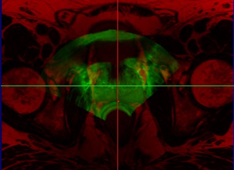
Prostate Fusion BiopsyTransrectal ultrasound (TRUS) guided biopsy remains the gold standard for diagnosis. However, it suffers from low sensitivity, leading to an elevated rate of false negative results. On the other hand, the recent advent of PET imaging using a novel dedicated radiotracer, Ga-labelled PSMA (Prostate Specific Membrane Antigen), combined with MR provides improved preinterventional identification of suspicious areas. Thus, MRI/TRUS fusion image-guided biopsy has evolved to be the method of choice to circumvent the limitations of TRUS-only biopsy. We propose a multimodal fusion image-guided biopsy framework that combines PET-MRI images with TRUS. Based on open-source software libraries, it is low cost, simple to use and has minimal overhead in clinical workflow. It is ideal as a research platform for the implementation and rapid bench to bedside translation of new image registration and visualization approaches. |
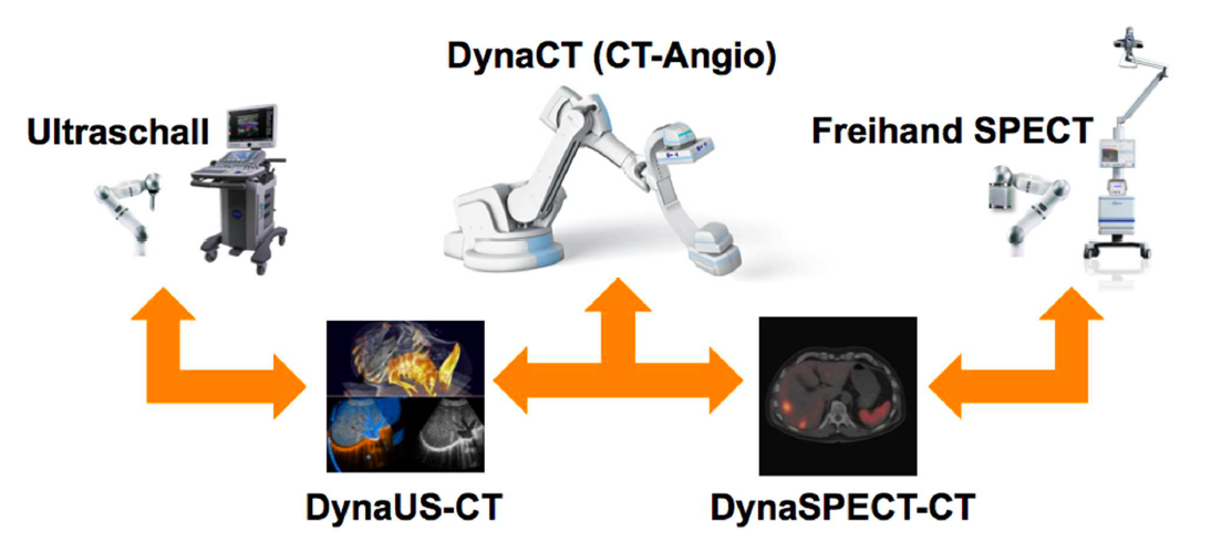
Advanced Robotics for Multi-Modal Interventional Imaging (RoBildOR)This project aims at developing advanced methods for robotic image acquisitions, enabling more flexible, patient- and process-specific functional and anatomical imaging with the operating theater. Using robotic manipulations, co-registered and dynamic imaging can be provided to the surgeon, allowing for optimal implementation of preoperative planning. In particular, this projects is the first one developing concepts for intraoperative SPECT-CT, and introduces intraoperative robotic Ultrasound imaging based on CT trajectory planning, enabling registration with angiographic data. With distinguished partners from Bavarian industry, this project has a fundamental contribution in developing safe, reliable, flexible, and multi-modal imaging technologies for the operating room of the future. |
SUPRA: Software Defined Ultrasound Processing for Real-Time ApplicationsSUPRA is an open-source pipeline for fully software defined ultrasound processing for real-time applications. Covering everything from beamforming to output of B-Mode images, SUPRA can help to improve the reproducibility of results and does allow for a full customization of the image acquisition workflow. Including all processing stages of a common ultrasound pipeline, it can be executed in 2D and 3D on consumer GPUs in real-time. Even on hardware as small as the CUDA enabled Jetson TX2, SUPRA allows for 2D imaging in real-time. You can access the code on our github page: https://github.com/IFL-CAMP/supra Additional information can be found in our work on SUPRAGöbl, R. and Navab, N. and Hennersperger, C., SUPRA: Open Source Software Defined Ultrasound Processing for Real-Time Applications, eprint arXiv:1711.06127, Nov 2017, under review for IPCAI2018The development of SUPRA was partly funded by the European Horizon 2020 Project EDEN2020. |
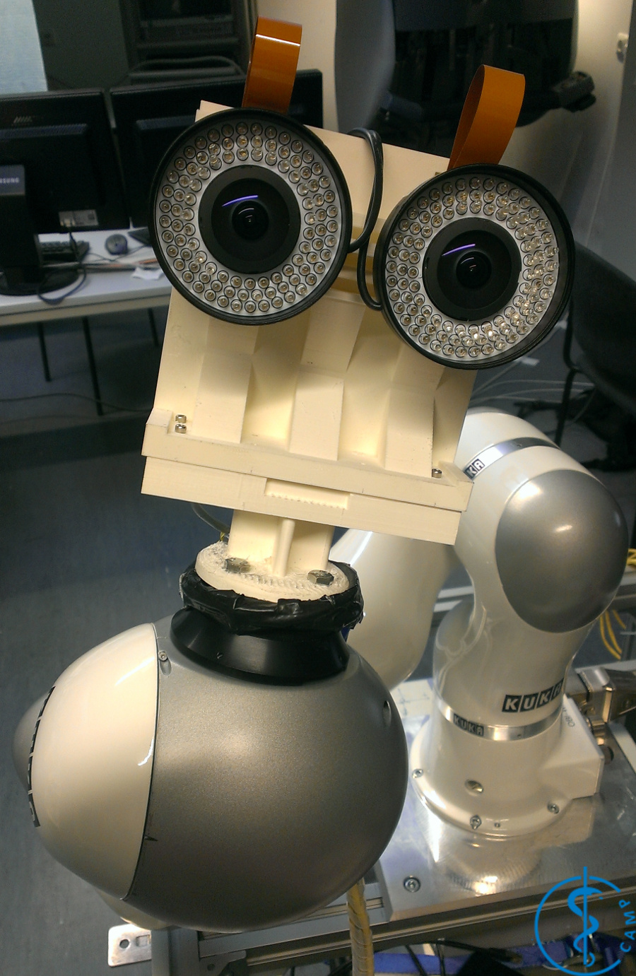
Inside-Out TrackingCurrent tracking solutions routinely used in a clinical, potentially surgically sterile, environment are limited to mechanical, electromagnetic or classic optical tracking. Main limitations of these technologies are respectively the size of the arm, the influence of ferromagnetic parts on the magnetic field and the line of sight between the cameras and tracking targets. These drawbacks limit the use of tracking in a clinical environment. The aim of this project is the development of so-called inside-out tracking, where one or more small cameras are fixed on clinical tools or robotic arms to provide tracking, both relative to other tools and static targets.These developments are funded from the 1st of January 2016 to 31st of December 2017 by the ZIM project Inside-Out Tracking for Medical Applications (IOTMA). |
EDEN2020: Enhanced Delivery Ecosystem for NeurosurgeryEDEN2020 (Enhanced Delivery Ecosystem for Neurosurgery) aims to develop the gold standard for one-stop diagnosis and treatment of brain disease by delivering an integrated technology platform for minimally invasive neurosurgery. A team of first-class industrial partners (Renishaw plc. and XoGraph ltd.), leading clinical oncological neurosurgery team (Università di Milano, San Raffaele and Politecnico di Milano) lead by Prof. Lorenzo Bello and the involvement of leading experts in shape sensing (Universitair Medisch Centrum Groningen) under supervision of Prof. Dr. Sarthak Misra The project is coordinated by Dr. Rodriguez y Baena, Imperial College London. His team provides the core technology for the envisioned system, the bendable robotic needle. During the course of EDEN2020 this interdisciplinary team will work on the integration of 5 key concepts, namely (1) pre-operative MRI and diffusion-MRI imaging, (2) intra-operative ultrasounds, (3) robotic assisted catheter steering, (4) brain diffusion modelling, and (5) a robotics assisted neurosurgical robotic product (the Neuromate), into a pre-commercial prototype which meets the pressing demand for better and less invasive neurosurgery. Our chair will be focusing on the imaging components (i.e. (1) and (2)), targeting the realtime compensation of tissue movement and accurate localization of the flexible catheters at hand. We will further extend the findings of FP7 ACTIVE, in which we successfully combined pre-operative MRI with intra-operative US through deformable 3D-2D registration, making us most qualified for this role. |
Computational Sonography3D ultrasound imaging has high potential for various clinical applications, but often suffers from high operator-dependency and the directionality of the acquired data. State-of-the-art systems mostly perform compounding of the image data prior to further processing and visualization, resulting in 3D volumes of scalar intensities. This work presents computational sonography as a novel concept to represent 3D ultrasound as tensor instead of scalar fields, mapping a full and arbitrary 3D acquisition to the reconstructed data. The proposed representation compactly preserves significantly more information about the anatomy-specific and direction-depend acquisition, facilitating both targeted data processing and improved visualization. We show the potential of this paradigm on ultrasound phantom data as well as on clinically acquired data for acquisitions of the femoral, brachial and antebrachial bone. Further investigation will consider additional compact directional-dependent representations on the one hand and on the other hand modify Computational Sonography from working on B-Mode images to RF-envelope statistics, motivated by the statistical process of image formation. We will show the advantages of the proposed improvements on simulated ultrasound data, phantom and clinically acquired ultrasound data. |
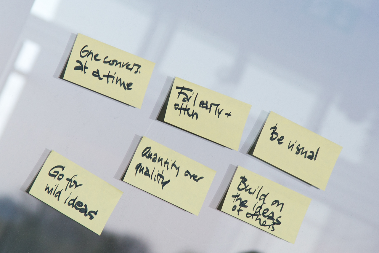
MedInnovate: From unmet clinical needs to solution conceptsLearn how to successfully identify unmet clinical needs within the clinical routine and work towards possible and realistic solutions to solve those needs. Students will get to know tools helping them to be successful innovators in medical technology. This will include all steps from needs finding and selection to defining appropriate solution concepts, including the development of first prototypes. Get introduced to necessary steps for successful idea and concept creation and realize your project in an interdisciplinary teams comprising of physicists, informations scientists and business majors. During the project phase, you are supported by coaches from both industry and medicine, in order to allow for direct and continuous exchange. |
| UsersForm | |
|---|---|
| Title: | Dr. |
| Circumference of your head (in cm): | |
| Firstname: | Christoph |
| Middlename: | |
| Lastname: | Hennersperger |
| Picture: |  |
| Birthday: | 1987/02/22 |
| Nationality: | Bavaria |
| Languages: | English, German |
| Groups: | Registration/Visualization, Segmentation, Reconstruction, Medical Imaging, Sensing, IFL, Ultrasound |
| Expertise: | Registration/Visualization, Segmentation, Medical Imaging, Computer-Aided Surgery, Sensing |
| Position: | Scientific Staff |
| Status: | Alumni |
| Emailbefore: | christoph.hennersperger |
| Emailafter: | tum.de |
| Room: | |
| Telephone: | |
| Alumniactivity: | Co-Founder and CTO at OneProjects? |
| Defensedate: | 19 June 2015 |
| Thesistitle: | Domain-Specific Modeling for Vascular Freehand Ultrasound |
| Alumnihomepage: | https://www.one-projects.com |
| Personalvideo01: | |
| Personalvideotext01: | |
| Personalvideopreview01: | |
| Personalvideo02: | |
| Personalvideotext02: | |
| Personalvideopreview02: | |
| I | Attachment  | Action | Size | Date | Who | Comment |
|---|---|---|---|---|---|---|
| | ch_gray_s.jpg | manage | 229.3 K | 07 Oct 2020 - 15:33 | ChristophHennersperger |
Edit |
Attach |
Refresh |
Diffs |
More |
Revision r1.38 - 16 Feb 2021 - 14:49 - ChristophHennersperger
