Keywords: Segmentation, Medical Imaging
Abstract
Development and refinement of a software system for semi-automatic segmentation of the patellar cartilage is the main goal of this project. By providing tools for sub-pixel accurate edge tracing, automatic contour completion, and adequate visualization, a remarkable speed-up of the physicians segmentation process can be achieved. Also, improved exactness can be reached for cartilage segmentation if expertise and automation are merged in a meaningful way.
Detailed Project Description
see also: Assessment of Knee Cartilage
Pictures

|
|
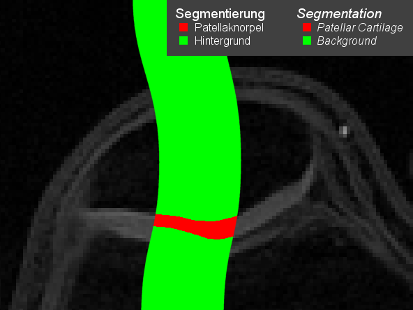
Figure 1: Part of the patellar cartilage segmentation (red vs. green background) superimposed onto original magnetic resonance image of a volunteer’s knee. On the lefthand side, there is no contrast between the patellar and femoral cartilages. Standard (contrast based) automatic segmentation algorithms will fail in such situations. On the righthand side, however, fully manual segmentation would be unnecessarily tedious.
|
|

|
|
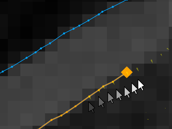
Figure 2: Tracing a contour. Holding the mouse button, the user drags to the right in this situation, roughly indicating the location of the cartilage surface (orange). The contour automatically sticks to the sub-pixel accurate egde points (small yellow wedges) computed in a preprocessing step.
|
|

|
|
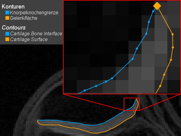
Figure 3: Complete segmentation of one slice. Magnification of a detail in the upper right corner reveals the sub-pixel accuracy of the segmentation.
|
|

|
|
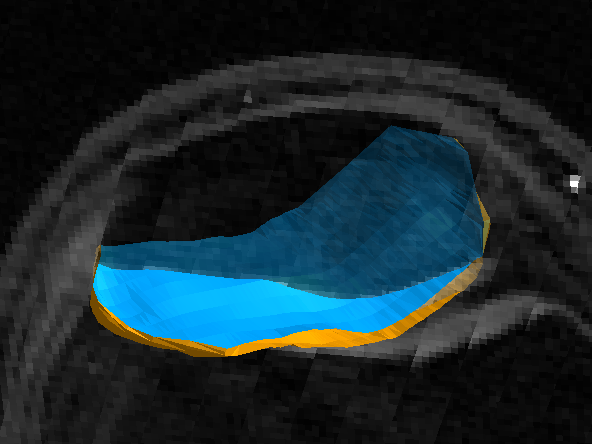
Figure 4: Combined rendition of skew plane of original MRI volume and segmented patellar cartilage.
|
|
Publications
| 2010 |
|
J. M. Raya Garcia del Olmo, A. Horng, L. König, M. Reiser, Ch. Glaser
Detecting Statistically Significant Changes in Cartilage Thickness with Sub-Voxel Precision
Proceedings of the 18th congress of the International Society for Magnetic Resonance in Medicine (ISMRM 2010), Stockholm, Sweden. Presentation 3192, electronic poster session “Meniscus & Cartilage”
(bib)
|
| 2007 |


|
L. König, M. Groher, A. Keil, Ch. Glaser, M. Reiser, N. Navab
Semi-Automatic Segmentation of the Patellar Cartilage in MRI
Proc. of Bildverarbeitung für die Medizin (BVM 2007), Munich, Germany, March 2007. The original publication is available online at www.springerlink.com.
(bib)
|
Team
Contact Person(s)
Working Group
Alumni
Location
 Visit our lab at Klinikum Grosshadern.
internal project page
Please contact
Ben Glocker for available student projects within this research project.


