| AMI-ARCS 2008, a one day satellite workshop of MICCAI 2008, will serve as a forum for researchers involved in all aspects of Augmented environments for Medical Imaging including Augmented Reality in Computer-aided Surgery. | 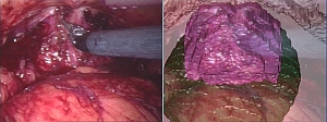 |
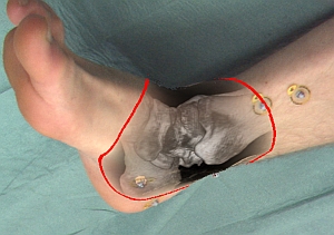 | In medical imaging, augmented environments aim to provide the physician with enhanced perception of the patient either by fusing various imaging modalities or by presenting image-derived information overlaid on the physician's view, establishing a direct relation between the image and the patient. The focus of this workshop will be innovative research into the technical aspects of augmentation, such as hardware and optical design, real-time implementations, registration and fusion, calibration, visualization and 3D perception as well as validation, clinical applications and clinical evaluation. In particular we will expect those presenting applications to describe the path to clinical introduction of their systems. We aim to include not only researchers in augmented reality but also those working on broader aspects of augmentation such as image fusion, simulation and rendering. AMI-ARCS will bring together researchers in computer science, electrical engineering, physics, and clinical medicine to present the state-of-the-art developments in this ever-growing research area. | |
| Dates | Workshop | Sept 10, 2008 in New York, USA 8am - 12pm, 1pm - 5pm |
| Call for papers | May 16th | |
| Submission deadline | June 16th | |
| Notification | July 21th | |
| Final camera ready version | August 7st | |
Proceedings online
Download the proceedings here!Program Overview
| 8.00 | Opening by general chairs |
| 8.10 | Session #1 (Applications) |
| 9.30 | Poster Session |
| 10.00 | Coffee break |
| 10.25 | Invited speaker - Professor Yoshinobu Sato |
| 11.15 | Panel Discussion |
| 11.45 | Lunch and demos |
| 12.45 | Session #2 (Accuracy & Assessment) |
| 14.05 | Invited speaker - Dr Mirna Lerotic |
| 14.55 | Coffee break |
| 15.10 | Session #3 (Display & Visualization) |
| 16.50 | Closing Remarks |
| 17.00 | Close |
Final Program
Download the full proceedings here!| 8.00 | Opening by general chairs | |
|---|---|---|
| 8.10 | Session #1 (Applications) | |
| 8.10 | A Training Suite for Image Overlay and Other Needle Insertion Techniques | Gregory S Fischer, Iulian Iordachita, Paweena U-Thainual, John A Carrino, Gabor Fichtinger |
| 8.30 | A 3D stereo system to assist surgical treatment of prostate cancer | Dongbin Chen, Erik Mayer, Justin Vale, Ann Anstee, Ara Darzi, Guang-Zhong Yang, Philip "Eddie" Edwards |
| 8.50 | Dr Gerard Guiraudon - Clinical preamble to the following two talks | |
| 9.00 | Where Cardiac Surgery Meets Virtual Reality: Pains and Gains of Clinical Translation | Christian A. Linte, Terry M. Peters |
| 9.15 | Accuracy of Tracked 2D Ultrasound during Minimally Invasive Cardiac Surgery | Andrew D. Wiles, Cristian A. Linte, Chris Wedlake, John Moore, Terry M. Peters |
| 9.30 | Poster Session | |
| Tracking-based statistical correction for radio-guided cancer surgery | Alexander Hartl, Thomas Wendler, Tobias Lasser, Sibylle I. Ziegler, Nassir Navab | |
| Registration of a 4D Cardiac Motion Model to Endoscopic Video for Augmented Reality Image Guidance of Robotic Coronary Artery Bypass | Michael Figl, Daniel Rueckert, David Hawkes, Roberto Casula, Mingxing Hu, Ose Pedro, Dong Ping Zhang, Graeme Penney, Fernando Bello, Philip Edwards | |
| A Method for Accelerating Bronchoscope Tracking Based on Image Registration by GPGPU | Takamasa Sugiura, Daisuke Deguchi, Takayuki Kitasaka, Kensaku Mori, Yasuhito Suenaga | |
| Semi-Automated Segmentation of Pelvic Organs for AR Guided Robotic Prostatectomy | Wenzhe Shi, Lin Mei, Dongbin Chen, Ara Darzi, Philip Eddie Edwards | |
| 10.00 | Coffee Break | |
| 10.25 | Invited speaker - Professor Yoshinobu Sato (see below) | |
| 11.15 | Panel Discussion | |
| 11.45 | Lunch and Demos | |
| 12.45 | Session #2 (Accuracy & Assessment) | |
| 12.45 | Workflow Based Assessment of the Camera Augmented Mobile C-arm System | Joerg Traub, Seyed-Ahmad Ahmadi, Nicolas Padoy, Lejing Wang, Sandro Michael Heining, Ekkehard Euler, Pierre Jannin, Nassir Navab |
| 13.05 | The Visible Korean Human Phantom: Realistic Test & Development Environments for Medical Augmented Reality | Christoph Paul Bichlmeier, Ben Ockert, Oliver Kutter, Mohammad Rustaee, Sandro Michael Heining, Nassir Navab |
| 13.25 | Benefits of augmented reality function for laparoscopic and endoscopic surgical robot systems | Naoki Suzuki, Asaki Hattori, Makoto Hashizume |
| 13.45 | Refining the 3D surface of blood vessels from a reduced set of 2D DSA images | Erwan Kerrien, Marie-Odile Berger, Jérémie Dequidt |
| 14.05 | Invited speaker - Dr Mirna Lerotic (see below) | |
| 14.55 | Coffee Break | |
| 15.10 | Session #3 (Display & Visualization) | |
| 15.10 | Contextually Enhanced 3D Visualization for Multi-burn Tumor Ablation Guidance | Andrei State, Hua Yang, Henry Fuchs, Sang Woo Lee, Patrick McNeillie, Charles Burke |
| 15.30 | Video to CT Registration for Image Overlay on Solid Organs | Balasz Vagvolgyi, Li-Ming Su, Russell Taylor, Gregory Donald Hager |
| 15.50 | Integrating Autostereoscopic Multi-View Lenticular Displays in Minimally Invasive Angiography | Daniel Ruijters |
| 16.10 | In-Situ Visualization of Medical Images Using Holographic Optics | John M. Galeotti, Mel Siegel, George Stetten |
| 16.30 | Real-time Volume Rendering for High Quality Visualization in Augmented Reality | Oliver Kutter, André Aichert, Christoph Bichlmeier, Joerg Traub, Sandro Michael Heining, Ben Ockert, Ekkehard Euler, Nassir Navab |
| 16.50 | Closing Remarks | |
| 17.00 | Close | |
Call for demos
- We offer the possibility to show live demos of your system
- We may offer stereoscopic visualization facilities in order to appreciate your visualization in 3D
Format of workshop
The format will be a full day workshop. The majority of the program will comprise innovative papers from researchers given as oral presentations. There will be a demonstration session during lunch, which will take place instead of a poster session, since augmented reality developments are not generally well presented by posters. There will also be a short discussion session at the end of the day. Anticipated number of participants: This workshop will be the third in a series of MICCAI workshops on augmented environments for medical imaging following AMI-ARCS 2004 & 2006. These had between 40 and 70 participants and this year we would anticipate attendance to be in a similar range.Submission
The submission deadline has been passed. Your reviews will be available at the submission site.Invited speakers
 | Professor Yoshinobu Sato, Division of Image Analysis, Osaka University, Japan, will give a talk entitled Deformation for endoscopic surgical navigation about his work in augmented reality for thoracic and abdominal surgery including clinical experience with his system. ABSTRACT: An important issue in clinical applications of endoscopic surgical navigation for the thoracic and abdominal organs is organ motion and deformation during surgery as well as between pre- and intraoperative patient postures. In this talk, we describe our recent efforts to address this issue from the three aspects; (1) intraoperative motion data acquisition using intra-body magnetic trackers and laparoscopic 3D ultrasound systems, (2) intraoperative deformation estimation by combining pre- and intra-operative data, and (3) intraoperative deformation prediction using anatomical prior information, that is, atlas-based approach. In addition to in vitro and in vivo animal experiments, clinical tests are demonstrated for thoracoscopic lung cancer resection and urology applications |   |
 | Dr Mirna Lerotic, Imperial College, London, UK, will speak about her work in augmented reality depth perception in a talk titled Augmented reality for minimally invasive surgery. ABSTRACT: Minimally Invasive Surgery (MIS) provides an ideal platform for using Augmented Reality (AR) for performing image guided surgery. A non-photorealistic rendering based AR technique for providing see-through vision of the embedded virtual object whilst maintaining salient anatomical details of the exposed anatomical surface will be presented. The method provides improved depth perception in stereo environments. | 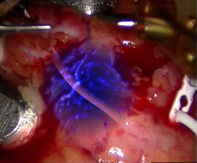 |
Committees
General Chairs
- PJ "Eddie" Edwards, Biosurgery and Surgical Technology Department, Imperial College, London UK
- Kensaku Mori, Department of Media Science, Graduate School of Information Science, Nagoya University
- Tobias Sielhorst (CAMP, TU Munich, Germany, )
Program Committee
- Wolfgang Birkfellner
- Stephane Nicolau
- Joerg Traub
- Nassir Navab
- Terry Peters
- Jannick Rolland
- Adrien Bartoli
- Marie-Odile Berger
- Rudy Lapeer
- George Stetten
- Ali Khamene
- Sandro Heining, MD