NaNuLab

|
NaNuLab stands for Navigated Nuclear Medicine Laboratory, a joint research laboratory of the Nuclear Medicine Department of the Klinikum rechts der Isar and the Chair for Computer Aided Medical Procedures of the Computer Science Department.
Address: Dr. Sibylle Ziegler / Thomas Wendler Nuklearmedizinische Klinik und Poliklinik Klinikum rechts der Isar Ismaninger Str. 22 81675 München Phone: +49 (89) 4140-2987 Fax: +49 (89) 4140-4950 Contact details and directions to NaNuLab can be found here. |
Facilities
Projects @ NaNuLab
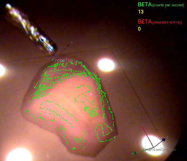
Navigated Beta Probes for Optimal Tumor Resectionin collaboration with Nuclear Medicine Department at Klinikum rechts der Isar In minimally invasive tumor resection, the goal is to perform a minimal but complete removal of cancerous cells. In the last decades interventional beta probes supported the detection of remaining tumor cells. However, scanning the patient with an intraoperative probe and applying the treatment are not done simultaneously. The main contribution of this work is to extend the one dimensional signal of a nuclear probe to a four dimensional signal including the spatial information of the distal end of the probe. This signal can be then used to guide the surgeon in the resection of residual tissue and thus increase its spatial accuracy while allowing minimal impact on the patient. |
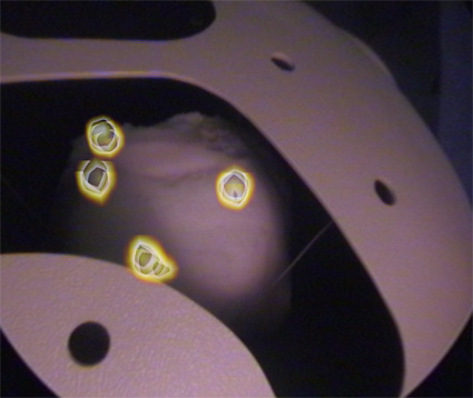
Navigated Gamma Probes for Tumor Localizationin collaboration with Nuclear Medicine Department at Klinikum rechts der Isar Nuclear medicine imaging modalities assist commonly in surgical guidance given their functional nature. However, when used in the operating room they present limitations. Pre-operative tomographic 3D imaging can only serve as a vague guidance intra-operatively, due to movement, deformation and changes in anatomy since the time of imaging, while standard intra-operative nuclear measurements are limited to 1D or (in some cases) 2D images with no depth information. To resolve this problem we propose the synchronized acquisition of position, orientation and readings of gamma probes intra-operatively to reconstruct a 3D activity volume. In contrast to conventional emission tomography, here, in a first proof-of-concept, the reconstruction succeeds without requiring symmetry in the positions and angles of acquisition, which allows greater flexibility and thus opens doors towards 3D intra-operative nuclear imaging. |


