Brain tumor segmentation and characterisationIn medical collaboration with:Klinikum rechts der Isar der TU München, Neuroradiologie: Prof. Dr. med. Claus Zimmer and Priv.-Doz. Dr. med. Jan Stefan Bauer Scientific Director: Prof. Dr. Bjoern Menze Contact Person(s): Esther Alberts |
Abstract
A generative-discriminative framework is built for multi-modal brain tumor segmentation with the use of longitudinal growth models. The framework will be validated and compared with the state-of-the-art. In a later stage, image features based on tumor-specific bio-markers are explored, and, more specifically, their contribution to more accurate brain tumor segmentation is studied.Detailed Project Description
Data acquisition and pre-processingWe use MR images of four modalities: T1, T1c, T2 and FLAIR. Images are pre-processed by means of ITK MR biasfield correction and NiftiReg non-rigid registrations.
Image segmentation techniques
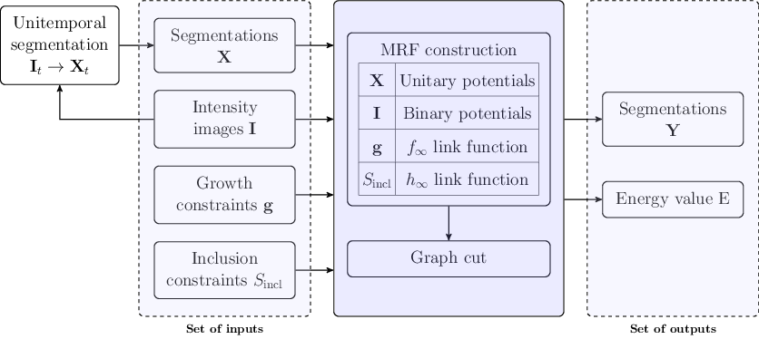
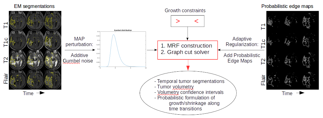
We use a generative tumor segmentation algorithm based on Expectation-Maximisation, which calculates spatial tumor probability maps for every modality. These tumor probability maps are subsequently spatially and temporally regularized using a 4-dimensional Markov random field solved by graph cut.
Patient-specific tumor growth estimations
A non-parametric growth model is build on top of the graph cut segmentations and estimates tumor volumes along time.
Pictures
Clinical Relevance
Oncology, NeuroradiologyTeam
Contact Person(s)
|
Working Group
|
|
Location
| Technische Universität München Institut für Informatik / I16 Boltzmannstr. 3 85748 Garching bei München Tel.: +49 89 289-17058 Fax: +49 89 289-17059 |
| Klinikum rechts der Isar der Technischen Universitüt München Ismaninger Str. 22 81675 München IFL Lab - Room: 01.3a-c Tel.: +49 89 4140-6457 Fax: +49 89 4140-6458 |
internal project page
Please contact Esther Alberts for available student projects within this research project.


