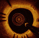
OCT Tissue ClassificationIn medical collaboration with:Stephane Carlier, Andreas Koenig and Adnan Kastrati, Deutsches Herzzentrum, Munich Scientific Director: Nassir Navab Contact Person(s): Sailesh Conjeti, Amin Katouzian , |
Abstract
Optical coherence tomography (OCT), employing light rather than ultrasound, is a high-resolution imaging technology that permits a precise assessment of biological tissue. Used intravascular, OCT is increasingly used for assessing safety and efficacy of intracoronary devices, such as drug-eluting stents and bioabsorbable stents. Obtained images provide insights regarding stent malposition, overlap, and neointimal thickening, among others. Recent OCT histopathology correlation studies have shown that OCT can be used to identify plaque composition, and hence it is possible to distinguish “normal” from “abnormal” neointimal tissue based on its visual appearance. The aim of this project is to develop a novel method for the automatic analysis of tissue in IVOCT. Automatic tissue classification will allow for a quantitative and potentially more time-efficient and objective analysis of IVOCT data. For instance, classifying neointimal tissue as either “mature” or “immature” can be used to assess the disease state of patients; as a potential predictor of late stent-failure events such as stent thrombosis and restenosis.Team
Contact Person(s)
|
|
Working Group
|
|
|
|
Location
| Technische Universität München Institut für Informatik / I16 Boltzmannstr. 3 85748 Garching bei München Tel.: +49 89 289-17058 Fax: +49 89 289-17059 |
| Deutsches Herzzentrum München Klinik an der Technischen Universität München Lazarettstr. 62 80636 München Room: 215 Tel.: +49 89 1812 3710 |
internal project page
Please contact Sailesh Conjeti, Amin Katouzian , for available student projects within this research project.


