Keywords: Segmentation
Abstract
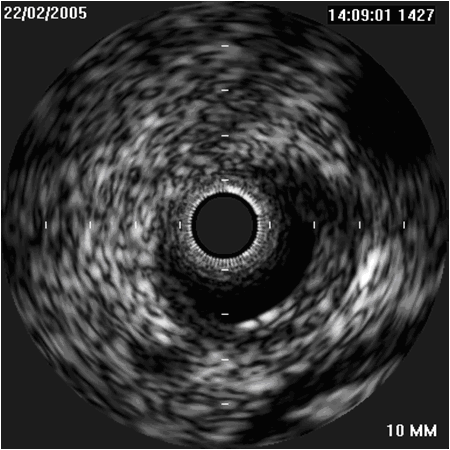
Heart attack and stroke are the major causes of human death and atherosclerotic plaques are the most common effect of cardiovascular disease. Intravascular ultrasound (IVUS), a diagnostic imaging technique, offers a unique view of the morphology of the arterial plaque and displays the morphological and histological properties of a cross-section of the vessel. Limitations of the grayscale IVUS manual plaque assessment have led to the development of quantitative techniques for analysis of characteristics of plaque components. In vivo plaque characterization with the so called Virtual Histology (VH)-IVUS, which is based on the ultrasound RF signal processing, is widely available for atherosclerosis plaque characterization in IVUS images. However, it suffers from a poor longitudinal resolution due to the ECG-gated acquisition. The focus of this
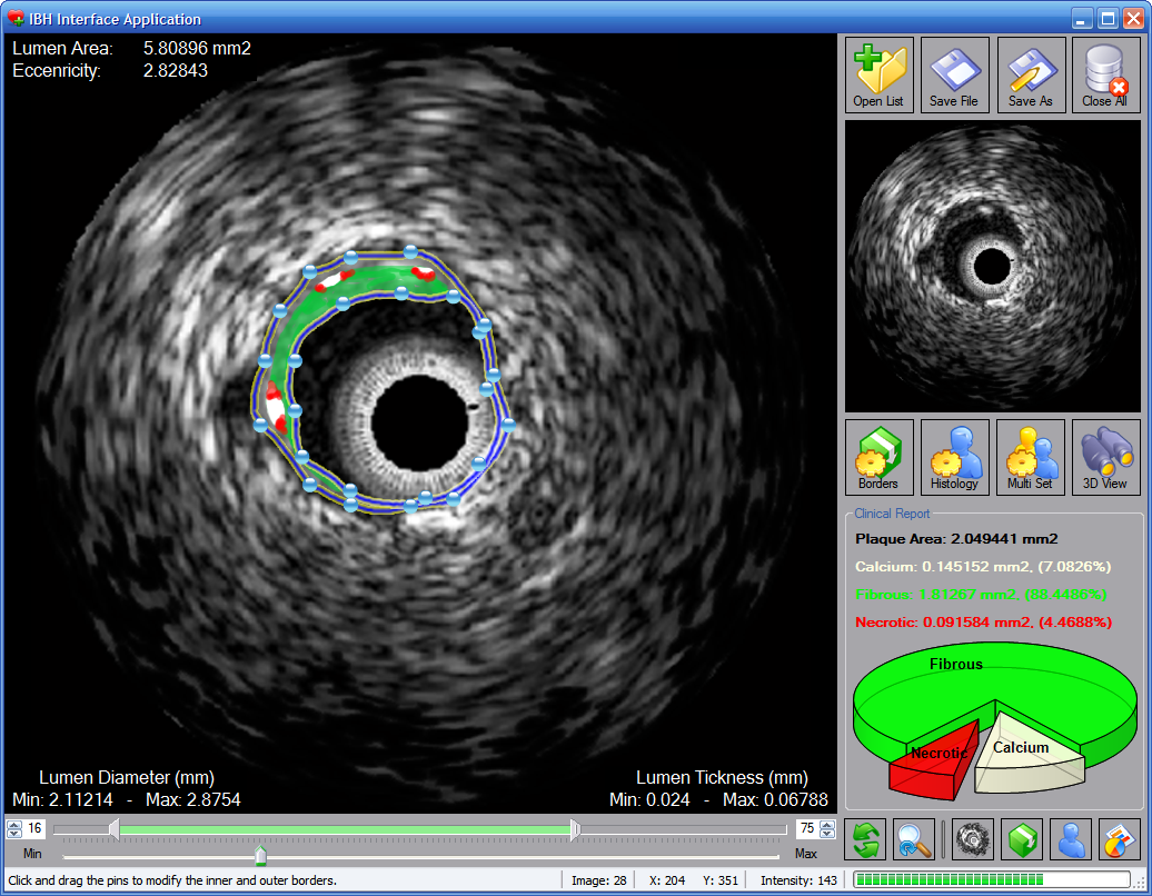
PhD? work is to provide effective methods for image-based vessel plaque characterization via IVUS image analysis to overcome the limitations of current techniques. The proposed algorithms are also applicable to the large amount of the IVUS image sequences obtained from patients in the past, where there is no access to the corresponding radio frequency(RF) data. Since the proposed method is applicable to all IVUS frames of the heart cycle, therefore it outperforms the longitudinal resolution of the so called VH method.
Pictures

|
|
Figure 1: IVUS Image
|
|

|
|
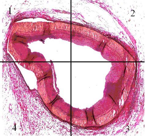
Figure 2: Histology
|
|

|
|
Figure 3: Manually Analyzed Image
|
|
Team
Contact Person(s)
Working Group
Location
internal project page
Please contact
Arash Taki for available student projects within this research project.


