Kooperationsprojekt SFB 824 (3. Förderperiode) & BFSIn medical collaboration with:Institut für Pathologie, Fakultät für Medizin, TUM Urologische Klinik und Poliklinik, Fakultät für Medizin, TUM Pathologisches Institut, Medizinischen Fakultät, LMU Urologische Klinik und Poliklinik, Medizinischen Fakultät, LMU Scientific Director: Prof. Dr. Nassir Navab, Dr. Katja Steiger, Dr. Maria Athelogou Contact Person(s): M.Sc. Beatrice Lentes and M.Sc. Iro Laina In industrial collaboration with: Definiens AG Morphisto GmbH |
Abstract
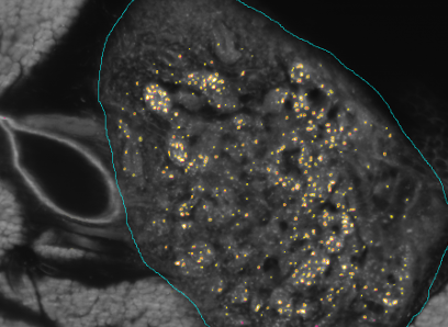
The SFB824 (Sonderforschungsbereich 824: Central project for histopathology, immunohistochemistry and analytical microscopy) represents an interdisciplinary consortium which aims at the development of novel imaging technologies for the selection and monitoring of cancer therapy as an important support for personalized medicine. Z2, the central unit for comparative morphomolecular pathology and computational validation, provides integration, registration and quantification of data obtained from both macroscopic and (sub-)cellular in-vivo as well as ex-vivo imaging modalities with tissue-based morphomolecular readouts as the basis for the development and establishment of personalized medicine. In order to develop novel imaging technologies, co-annotation and validation of image data acquired by preclinical or diagnostic imaging platforms via tissue based quantitative morphomolecular methods is crucial. Light sheet microscopy will continue to close the gap between 3D data acquired by in-vivo imaging and 2D histological slices especially focusing on tumor vascularization. The Multimodal ImagiNg Data Flow StUdy Lab (MINDFUL) is a central system for data management in preclinical studies developed within SFB824. Continuing the close collaboration of pathology, computer sciences and basic as well as translational researchers from SFB824 will allow the Z2 to develop and subsequently provide a broad variety of registration and analysis tools for joint imaging and tissue based image standardization and quantification.The goal of the BFS Project: ImmunoProfiling using Neuronal Networks (IPN2) is to develop a method based on neuronal networks and recent advances in Deep Learning to allow characterization of a patient's tumor as ″hot″ or ″cold″ tumor depending on the identified ImmunoProfile. Recent research has shown that many tumors are infiltrated by immuno-competent cells, as well as that the amount, type and location of the infiltrated lymph nodes in primary tumors provide valuable prognostic information. In contrast to a ″cold tumor″, a ″hot tumor″ is characterized by an active immune system which the tumor has identified as threat. This identification provides the basis for selecting the therapy best suitable for the individual patient.
Team
Contact Person(s)
|
|
Working Group
|
|
Location
| Klinikum rechts der Isar der Technischen Universitüt München Ismaninger Str. 22 81675 München IFL Lab - Room: 01.3a-c Tel.: +49 89 4140-6457 Fax: +49 89 4140-6458 |
internal project page
Please contact M.Sc. Beatrice Lentes and M.Sc. Iro Laina for available student projects within this research project.


