Image Guided Surgeries - From Bench to Bedside and Back
Summer term 2015
Administrative InfoLecture: Dr. Jörg Traub and Prof. Dr. Michael FriebeTutor: Philipp Matthies Type: Lecture + Lab Courses (Module IN2286) Programs: Informatics (Bachelor, Master) Biomedical Computing (Master) Information Systems (Wirtschaftsinformatik) (Bachelor, Master) SWS: 2+2 ECTS: 6 Credits Course Language: English |
Time, Location & RequirementsLecture: The lecture will be held in 4 blocks of six hours each (9:00am to 12:00am and 1:00pm to 4:00pm), the dates are:
Locations: Conference room in IAS basement and MI 03.13.010 We will have teams working on NOTES procedures, conventional laparoscopy / bronchoscopy, arthroscopy, stenting interventions, and possibly other. The surgeries will be performed in Munich hospitals. The teams will be asked to contact the head surgeons for an appointment and arrange for a Q&A session before and/or after the surgery. The surgery should be well documented -- please use an engineering approach to analyze the procedures. Please note that the lecturers will be available 30 minutes prior to and 1 hour after the lecture in their office in the TUM-IAS building (Room 2.004), 2nd floor, right, last one on the left. Requirements:
|
||||||||
Announcements
|
Photos
|
Lecture Schedule of Block 1 - April 17, 2015
| Time | Lecturer | Content | Material |
|---|---|---|---|
| 09:00 - 09:30 | Local: Dr. Jörg Traub | Introduction, Course Outline and Group Building | none |
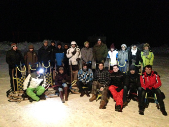
| 09:30 - 10:00 | Video conferencing: Dr. Jörg Traub and Prof. Dr. Michael Friebe (from Magdeburg) | Introduction to the Lecturers & Final Lecture Block with trip to a mountain area | |
| 10:10 - 11:30 | Video conferencing: Dr. Jörg Traub | OR Visits Preperation and Medical Ethics | |
| 10:30 - 11:15 | Video conferencing: Prof. Dr. Michael Friebe (from Magdeburg) | Motivation & Introduction in Medical Imaging | |
| 11:15 - 12:00 | Video conferencing: Dr. Jörg Traub | Motivation and Introduction in Medical Image Processing and Navigation | |
| 12:00 - 13:00 | lunch break | ||
| 13:00 - 16:00 | Local: Dr. Jörg Traub | Applications of IGS and OR Video Analysis |
Lecture Schedule of Block 2 - May 22, 2015
| Time | Lecturer | Content | Material |
|---|---|---|---|
| 09:00 - 10:00 | Video conferencing: invited presentation of Dr. Martin Groher from MicroDimensions GmbH | Applications of Image Processing in Pathology and the Creation of MicroDimensions GmbH | |
| 10:00 - 10:15 | Organizational & OR codex | | |
| 10:30 - 12:00 | Video conferencing: Dr. Jörg Traub (from Magdeburg) | Image Processing & Navigation in a Nutshell | |
| 12:00 - 12:45 | lunch break | ||
| 12:45 - 04:00 | Local: Prof. Dr. Michael Friebe | Imaging Systems I & II | |
Lecture Schedule of Block 3 - June 19, 2015
| Time | Lecturer | Content  | Material |
|---|---|---|---|
| 15:45 - 16:00 | Local: All | Q&A – project group work for final presentation and documentation | |
| 14:45 - 15:15 | Video conferencing: Dr. Jörg Traub | Project Management | |
| 09:00 - 12:00 | Local: Dr. Jörg Traub and every team | Midterm Presentation of OR Visits | |
| 15:15 - 15:45 | Video conferencing: Prof. Dr. Michael Friebe (from Magdeburg) | Innovation Games / Conceptual Blockbusting | |
| 14:00 - 14:30 | Video conferencing: invited presentation from Stefan Wiesner from HumediQ GmbH | Imaging and Guidance in Radiation Therapy | |
| 13:00 - 14:00 | Video conferencing: invited presentation of Dr. Hans Kiening from Arri Medical | Digitalization of Operation Microscope | |
| 14:30 - 14:45 | Video conferencing: Iurie Tap | A perspective from an alumnus of "Medical Technology Entrepreneurship" | |
| 12:00 - 13:00 | lunch break |
Lecture Schedule of Block 4 - July 16-18, 2015
| Time | Lecturer | Content | Material |
|---|---|---|---|
| German Healthcare System Overview | |
Readings
- D. Buckland, “How Surgeons and Engineers Can Communicate Better,” MedGadget, 08-Jul-2012. [Online]. Available: http://www.medgadget.com/2012/08/how-surgeons-and-engineers-can-communicate-better.html. [Accessed: 03-Apr-2015].
- T. Peters and K. Cleary, Image-Guided Interventions: Technology and Applications, Auflage: 2008. New York: Springer, 2008.
- F. L. Greene and B. T. Heniford, Minimally Invasive Cancer Management, 2nd ed. 2010. New York: Springer, 2010.
- T. M. Peters, “Image-guidance for surgical procedures,” Physics in Medicine and Biology, vol. 51, no. 14, pp. R505–R540, Jul. 2006.
- Ziv Yaniv and Kevin Cleary, “Image-Guided Procedures: A Review,” Georgetown University, CAIMR TR-2006-3, Apr. 2006.
- J. K. Udupa, V. R. LeBlanc, Y. Zhuge, C. Imielinska, H. Schmidt, L. M. Currie, B. E. Hirsch, and J. Woodburn, “A framework for evaluating image segmentation algorithms,” Computerized Medical Imaging and Graphics, vol. 30, no. 2, pp. 75–87, Mar. 2006.
- N. Ayache, O. Clatz, H. Delingette, G. Malandain, X. Pennec and M. Sermesant, “Asclepios : a Research Project-Team at INRIA for the Analysis and Simulation of Biomedical Images,” 2006.
- K. Doi, “Current status and future potential of computer-aided diagnosis in medical imaging,” British Journal of Radiology, vol. 78, no. suppl_1, pp. S3–S19, Jan. 2005.
- J. V. Hajnal, D. L. G. Hill, and D. J. Hawkes, Medical Image Registration. Boca Raton: Crc Pr Inc, 2001.
- Gary Bishop, Greg Welch, and B. Danette Allen, “Tracking: Beyond 15 Minutes of Thought - SIGGRAPH 2001 Courses - Course 11.” 2001.
- K. Cleary and C. Nguyen, “State of the art in surgical robotics: Clinical applications and technology challenges,” Computer Aided Surgery, vol. 6, no. 6, pp. 312–328, 2001.
- J. B. A. Maintz and M. A. Viergever, “An Overview of Medical Image Registration Methods,” In Symposium of the Belgian hospital physicists association (SBPH-BVZF, 1996.
- Ron Kikinis, David Altobelli, Langham Gleason, and Ferenc Jolesz, “Enhancing Reality in the Operating Room,” Brigham and Womens Hospital, Boston, 1993.
- A. Wolbarst and W. Hendee, Evolving and experimental technologies in medical imaging, Radiology, 238 (2006), pp. 16-39
- PhD Thesis - Chapter 1 & 2 - Joerg Traub, New Concepts for Design and Workflow Driven Evaluation of Computer Assisted Surgery Solutions, 2008, TUM, Munich, Germany
- Selected Workshop and Proceedings of the following congresses: IPCAI/CARS, MICCAI, SMIT
- POWER CARTOON ACCESS -- FOR THE PROJECT: Manuals downloadable at http://www.powtoon.com -- Student Access through here
| TeachingForm | |
|---|---|
| Title: | Image Guided Surgeries - From Bench to Bedside and Back |
| Professor: | Dr. Joerg Traub, Prof. Dr. Michael Friebe |
| Tutors: | Philipp Matthies |
| Type: | Lecture |
| Information: | 2 + 2 SWS, 6 ECTS Credits (Module IN2286) |
| Term: | 2015SoSe |
| Abstract: | Participants learn the basics of medical applications in minimally invasive image guided surgery and related hybrid imaging. They gain the knowledge to understand the complex medical environment as well as its challenges in order to improve these procedures (including logistics and imaging in the operating theatre of the future). With knowledge about technology and algorithm they can model and master complex solutions. Besides the medical and technical knowledge, definition of clinical, technical projects in the field of intra operative imaging and navigation for minimal invasive surgery is the core focus during the course. Exposure to real world problems makes this a unique setup for modeling and implementing creative solutions. |


