Navigated RF Ablation
Advisor: Nassir NavabSupervision by: Marco Feuerstein
Abstract
Currently, RF ablation needles are used to treat tumors in every part of the body. In order to place the tip of the needle in the center of the lesion, US imaging is commonly used for guidance. In this case, the surgeon tries to visualize the tumor and the needle in the same US image. The needle however is not visible in the US image before insertion, making it difficult to imagine the tumor location relative to the needle tip and orientation. Additionally, the surgeon needs to precisely handle the US transducer and the needle at the same time.The navigation system developed for this purpose as part of the CAMPAR framework helps to simplify the RF needle placement by dividing it into two simple consecutive tasks: lesion finding and needle placement. This way, the surgeon does not have to simultaneously hold the US probe and RF needle. Additionally, the needle can always be visualized relatively to the US plane.
NeedlePlacement application
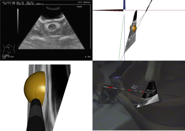
The ultrasound guided needle placement application helps a surgeon to insert the tip of a RF needle into a precise point, defined by the user. The system provides the means to calibrate the needle with high accuracy. It has four different views:Ultrasound view: The user sees the ultrasound video, with the option to freeze an image and define a spherical target. Using the ultrasound calibration results, the system maps the defined target to the world coordinate system, defined by the tracking device.
World interactive view: The surgeon can see the needle and the sphere target in a virtual environment. This view allows to specify the point of view using translation, rotation, and scale primitives. The model includes a line extending the needle and a line from its tip to the center of the target. The needle should be set in the direction where these two lines meet. One problem with this view is the need of mouse interaction, which is usually not practical.
Needle view: The user can see the needle and the target, in the direction of the needle. This is especially intuitive for directing the needle.
Augmented camera view: This view includes a camera video. The camera, like the US probe and the needle, is tracked and calibrated, so it is possible to super-impose the virtual models on top of the video. This way, the point of view is defined by the camera. The main advantage of this view is the ability to integrate other, non tracked elements into the environment. It also helps to qualitatively evaluate the accuracy of the system.
Screenshots

Literature
G. Scott Gazelle, S. Nahum Goldberg, Luigi Solviati and Tito Livraghi. Tumor Ablation with Radio-frequency Energy, in Radiology, 2000; 217, pp 633-646.
P-W. Hsu, R. W. Prager, A. H. Gee and G. M. Treece. Rapid, Easy and Reliable Calibration for Freehand 3D Ultrasound, in Ultrasound Med. Biol., June 2006; 32, pp 823-835.
C. Alcérreca, F. Arámbula and E. Hazan. Navigation system for computer assisted tumor ablation using radio frequency. IEEE - EMBC, NY, Sep 2006.
F. Sauer, A. Khamene and S. Vogt. An Augmented Reality Navigation System with a Single-Camera Tracker: System Design and Needle Biopsy Phantom Trial. MICCAI, 2002, Springer, Berlin, LNDC 2489, pp. 116-124
| ProjectForm | |
|---|---|
| Title: | Navigated RF Ablation |
| Abstract: | Currently, RF ablation needles are used to treat tumors in every part of the body. In order to place the tip of the needle in the center of the lesion, US imaging is commonly used for guidance. In this case, the surgeon tries to visualize the tumor and the needle in the same US image. The needle however is not visible in the US image before insertion, making it difficult to imagine the tumor location relative to the needle tip and orientation. Additionally, the surgeon needs to precisely handle the US transducer and the needle at the same time. The navigation system developed for this purpose as part of the CAMPAR framework helps to simplify the RF needle placement by dividing it into two simple consecutive tasks: lesion finding and needle placement. This way, the surgeon does not have to simultaneously hold the US probe and RF needle. Additionally, the needle can always be visualized relatively to the US plane. |
| Student: | Claudio Alcérreca |
| Director: | Nassir Navab |
| Supervisor: | Marco Feuerstein |
| Type: | Master Thesis |
| Status: | finished |
| Start: | 2006/07/01 |
| Finish: | 2006/10/27 |