User interface development and evaluation of a navigation system for bronchoscopy
Type: IDP with minor medicine, or SEPProject by: Arne Wirtz
Advisor: Nassir Navab, Hubert Hautmann
Supervision by: Tobias Reichl
Abstract
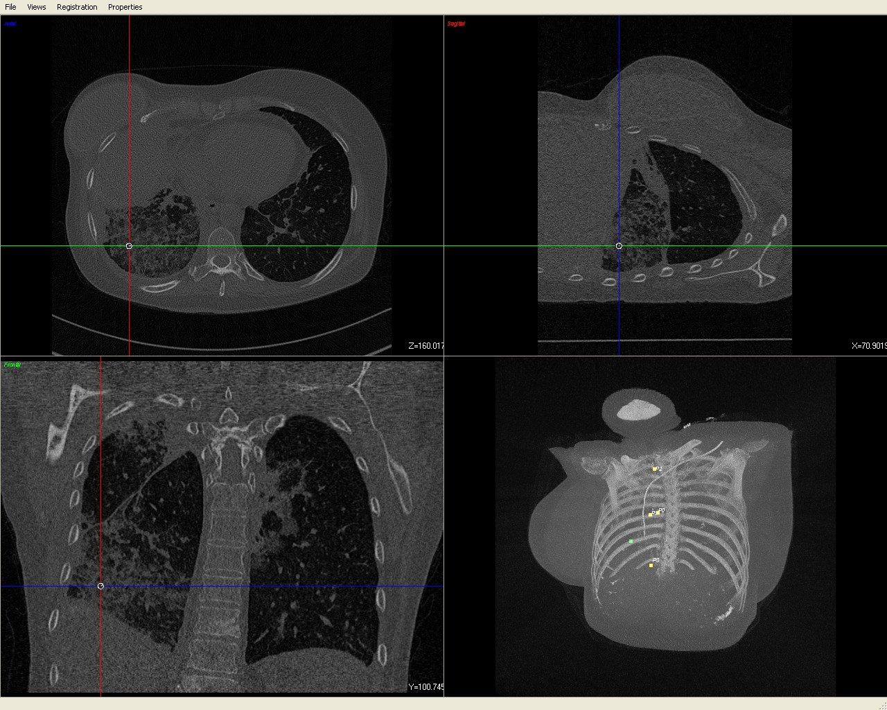
In bronchoscopy a physician examines the bronchial tree of a patient with a camera and medical instruments. To access regions outside the view of the camera, the only feedback of the position of the instrument is x-ray imaging. In these x-ray images the final lesion is not visible. The preoperative CT scan plus a electromagnetical tracking device is used to provide feedback of the current instrument location in real time. The work will be conducted in the pneumology department of the Klinikum rechts der Isar, Max-Weber-Platz. A first fully functional and tested prototype was developed by Julian Much based on the software framework CAMPAR. This software is based on C++, OpenGL and QT. The work will be in refining the user interface for navigation purposes and conduct together with Dr. Hautmann clinical experiments.
Task
Extension of the existing navigation software and user interface and clinical experiments (for more details see abstract below or live demo at Klinikum rechts der Isar).Requirements
Basic knowledge of C++ required. Knowledge of OpenGL and QT is of advantage.| Students.ProjectForm | |
|---|---|
| Title: | User interface development and evaluation of a navigation system for bronchoscopy |
| Abstract: | In bronchoscopy a physician examines the bronchial tree of a patient with a camera and medical instruments. To access regions outside the view of the camera, the only feedback of the position of the instrument is x-ray imaging. In these x-ray images the final lesion is not visible. The preoperative CT scan plus a electromagnetical tracking device is used to provide feedback of the current instrument location in real time. The work will be conducted in the pneumology department of the Klinikum rechts der Isar, Max-Weber-Platz. A first fully functional and tested prototype was developed by Julian Much based on the software framework CAMPAR. This software is based on C++, OpenGL and QT. The work will be in refining the user interface for navigation purposes and conduct together with Dr. Hautmann clinical experiments. |
| Student: | Arne Wirtz |
| Director: | Nassir Navab, Hubert Hautmann |
| Supervisor: | Tobias Reichl |
| Type: | IDP |
| Area: | Registration / Visualization, Computer-Aided Surgery |
| Status: | finished |
| Start: | 2009/04/01 |
| Finish: | 2010/03/09 |