Segmentation of Liver and Stroke Lesions in CT Scans via Phase Field Separation
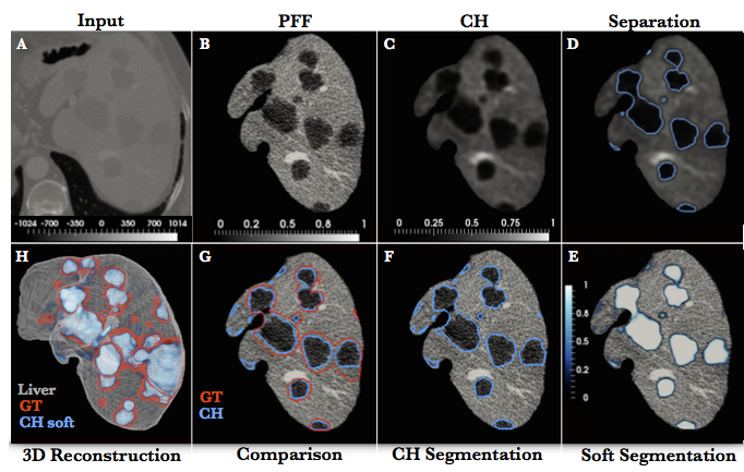
 Figure 1: A workflow of phase separation and lesions segmentation. A) cropped liver CT scan, converted into a phase field function (PFF) B), is used as initial condition for the Cahn-Hilliard (CH) equation, with the solution shown on C). D) the hard separation of the phases (blue line), is converted into a soft probabilistic segmentation E). F) the final hard segmentation (blue) in comparison with the ground truth (GT) (red) G). H) comparison of the GT (red) and soft segmentation (white-blue colormap) in a 3D representation.
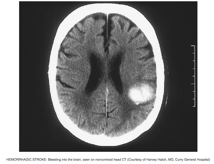
 Figure 2: CT scan of ischemic brain stroke.
|
Project Description
Segmentation of lesion is crucial for detection, diagnosis and monitoring disease progression. However, design of accurate automated methods remains challenging due to high noise in CT scans, low contrast between tissue and lesions, as well as large lesion variability. We propose a 3D automatic, unsupervised method for lesions segmentation using a phase separation approach. It is assumed that tissue is a mixture of multiple phases (e.g. healthy tissue and lesions) represented by different image intensities polluted by noise. The Cahn-Hilliard equation is used to remove the noise and separate the mixture into distinct phases (classes) with well-defined interfaces. This simplifies the lesion detection and segmentation task drastically and enables to segment lesions by thresholding the Cahn-Hilliard solution.
The Cahn-Hilliard separation (CHS) method was applied to the Liver Lesions Segmentation Challenge (LITS), where it ranked as the best unsupervised method. In this scenario liver tissue is consider as a mixture of two classes, i.e. healthy liver and lesions and CHS is applied to separate the system in two distinguish classes (see Figure 1). To further improve segmentation scores, a discriminative classifier can be trained to distinguish lesions and potential segmentation artifacts. The successful classifier with CHS approach will be applied to MICCAI Liver Lesions Segmentation Challenge this Autumn. To further exploit potential of the method to separate volume into multiple phases (classes), the CHS method will be applied to CT scan of stroke patient, where the system will be separated in three classes: healthy tissue (white and gray matter), cerbospinal fluid and lesion. The method can be applied to Stroke segmentation challenge in MICCAI conference this Autumn.
Tasks
- Train a discriminative classifier to further improve results of the liver lesion segmentation
- Extend and apply the CHS method to segmentation of stroke lesions (i.e. allow separation in three classes)
- Apply CHS method to the MICCAI challenge(s).
Requirements
- Good programming skill (C++, Python, Matlab)
- Basics of Numerical Analysis (not required but beneficial)
- Basic knowledge about classifiers and image processing
- Desire to learn and improvise
- Independent worker
Contact
If you are interested in the project or if you have any questions please contact
|
|
 Figure 1: A workflow of phase separation and lesions segmentation. A) cropped liver CT scan, converted into a phase field function (PFF) B), is used as initial condition for the Cahn-Hilliard (CH) equation, with the solution shown on C). D) the hard separation of the phases (blue line), is converted into a soft probabilistic segmentation E). F) the final hard segmentation (blue) in comparison with the ground truth (GT) (red) G). H) comparison of the GT (red) and soft segmentation (white-blue colormap) in a 3D representation.
 Figure 2: CT scan of ischemic brain stroke.