IDP: MR-Based Attenuation Correction for PET
Student: LorenSchwarzAdvisors: NassirNavab, Marc Huisman, Sibylle Ziegler, Maria Jose Martinez
Supervision by: AndreasKeil
Due date:
Abstract
A method is developed for PET attenuation correction based on MR images. MR scans that have been obtained using different sequences (T1, T2*, PD) are segmented into classes representing types of tissue. As an initial step, while performing experiments on mice, binary segmentation in the two classes air and tissue is considered sufficient. For later applications a distinction of air, bone and soft tissue is desired. After identifying the tissue type for each location within an MR image, an attenuation map is computed by assigning pre-defined attenuation coefficients to the segmented MR image. This attenuation map can then be used by PET scanners for attenuation correction.Motivation
Positron Emission Tomography (PET) is an important medical imaging modality that can provide an insight into the function of many human and animal organs. While in other modalities, such as Computed Tomography (CT) and Magnetic Resonance Imaging (MRI), mainly anatomical structures are visible, PET images show biochemical and physiological activity. Examination of small animals, such as mice and rats, is of great importance to biologists and to the pharmaceutical industry, where pre-clinical experiments are performed on small animals.Problem Statement
A crucial issue related to PET is the so-called attenuation effect. It is caused by absorption or diversion of photons by different kinds of tissue that they pass through on their way to the photon detectors. The location where photons are registered on the detectors is used to re-compute the position in space, where the photons originated from. Due to the attenuation effect, this calculated position will be erroneous. As a result, uncorrected PET images are blurred and have low contrast. The attenuation effect can, however, be corrected by using prior knowledge about attenuation properties of different tissue types. If it is known which tissues are being scanned by a PET system, the resulting PET image can be corrected. One common way to perform attenuation correction is to combine a PET scan with a CT scan. The two data sets are spatially registered and attenuation values for each location are calculated from the CT image. This is possible, since CT technology is based on X-rays which are being attenuated by different types of tissue. Mainly because of the side-effects of X-rays that are harmful to animals and humans, there is a great interest in using MRI technology for attenuation correction. This imaging modality, however, is based on totally different physical effects than CT imaging, so that there is no straightforward way of deriving attenuation values from an MR image. Moreover, MR scans of even identical anatomical structures can be highly different, depending on a large number of technical parameters.Project Procedures
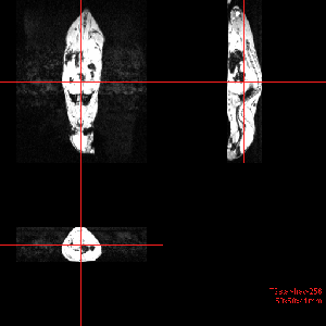
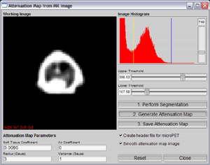
The outcome of this project was not foreseeable in its entirety from the beginning. Several important decisions depended on variables to be clarified in experiments. The procedures completed so far include the following: MR scans of mice in several sequences. The initial step was to perform MR scans of mice in order to find out what level of detail can be achieved with the available MR scanner (Figure 1). Best results have been obtained using the T1 and T2* sequences, where the former sequence showed best soft tissue contrast and the latter sequence showed bone structures more clearly. A special small animal coil was used in combination with a Philips human MR scanner. Evaluation of MR scans. Despite the high spatial resolution of the MR mice images, the level of detail showed to be hardly sufficient for segmentation into more than two classes. Bone structures are tiny - rarely larger than a few pixels - and moreover gray levels are obviously not linearly distributed over different types of tissue. In particular this means that a simple attribution of certain gray levels to specific tissue types (as it is done for CT images) is not possible. The only feasible approach without using advanced techniques, such as atlas based registration, seemed to be a simple binary segmentation into air and tissue. Acquisition of a whole PET-CT-MR data set. The idea was to compare attenuation maps obtained from a CT scan and a sample attenuation map generated from a binary segmented MR image. Due to technical problems with the PET and CT scanners this task could not be completely accomplished. However, another set of MR scans was performed, this time using a conventional human wrist coil instead of the specialized small animal coil. Image quality was not distinguishable between the two MR data sets. Implementation of a software module. According to nuclear medicine experts, the quality of the sample attenuation maps generated from MR images using a binary segmentation was better than what was achievable by a PET transmission scan for small animals. Therefore a software module was implemented for practical use with further small animal experiments. The module allows to set thresholds in order to obtain a binary segmentation of an MR scan. Attenuation coefficients can then be entered and assigned to the two resulting classes. Attenuation maps can be exported in a data format suitable for the microPET small animal PET scanner (Figure 2). Problem discussion. Given the difficulties in segmenting more than two classes from MR scans, the most important question became to evaluate how important a more advanced segmentation is in practice. In other words: would there be a significant negative influence on PET image correction, if an attenuation map was used that does not distinguish between soft tissue and bone? According to specialists at the nuclear medicine lab (and Chow et al.), this negative influence is negligible for small animal imaging. Therefore the decision was made to shift focus to human clinical data. Work on human clinical data. A twofold approach has been developed in order to assess the quality of attenuation maps that can be obtained by binary segmentation. On the one hand, CT-based attenuation maps are examined to find out how detailed an attenuation map should be for practical use. CT-based attenuation maps are increasingly quantized to fewer intensity classes to approach characteristics of potential MR-based attenuation maps. On the other hand, a comparison of CT-based and MR-based attenuation maps is performed on two levels: the maps are directly compared using a similarity measure and the results of attenuation correction on a PET image are compared using the two attenuation maps.Images
Figure 1: MR scan of a mouse (T1 weighted). Figure 2: Dialog for attenuation map generation from MR scans using binary segmentation.

Literature
- Zaidi, H., Montandon, M.-L., Slosman, D.: Magnetic Resonance Imaging-guided Attenuation and Scatter Corrections in Three-dimensional Brain Positron Emission Tomography, Med. Phys., Volume 30, pp. 937-948; 2003
- Zaidi, H. and Hsegawa, B.: Determination of the Attenuation Map in Emission Tomography, J. Nucl. Med., Volume 44, pp. 291-315; 2003
- Chow, P. L. and Rannou, F. R.: Attenuation Correction for Small Animal PET tomographs, Phys. Med. Biol., Volume 50, pp. 1837-1850; 2005
- Rappoport, V., Carney, J. P., Townsend, D. W.: CT Tube-voltage Dependent Attenuation Correction Scheme for PET/CT Scanners, IEEE; 2004
- Burger, C. Goerres, G. et al.: PET Attenuation Coefficients from CT Images: Experimental Evaluation of the Transformation of CT imto PET 511-keV Attenuation Coefficients, European Journal of Nuclear Medicine, Volume 29, pp. 923-927; 2002
Materials
- IDP-final-pres.pdf: Final Presentation
- final.zip: Project Report Files
| Students.ProjectForm | |
|---|---|
| Title: | MR-Based Attenuation Correction for PET |
| Abstract: | |
| Student: | Loren Schwarz |
| Director: | Nassir Navab, Marc Huisman |
| Supervisor: | Andreas Keil |
| Type: | IDP |
| Status: | finished |
| Start: | 2006/03/01 |
| Finish: | 2007/06/15 |