IVUSSimulation
Intravascular Ultrasound Simulation from HistologyIn medical collaboration with:Department of Radiology - Klinikum Rechts der Isar Scientific Director: Prof. Dr. Nassir Navab Contact Person(s): Silvan Kraft |
Abstract
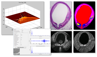
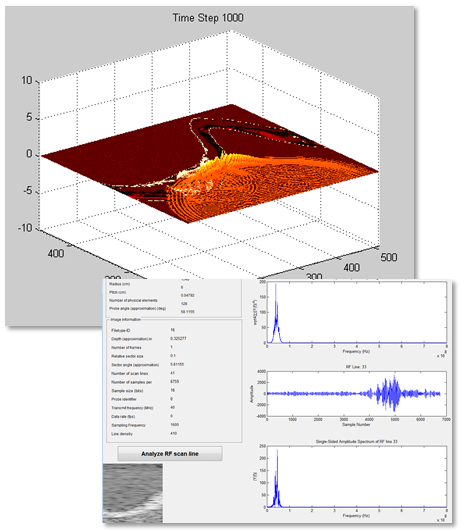
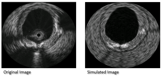
We introduce a framework to simulate intravascular ultrasound (IVUS) from histological sections. These sections were previously acquired along with real IVUS radiofrequency signals using single-element 40MHz transducer. After labeling and registering the section to the corresponding IVUS image, a virtual phantom was created, incorporating nuclei scatterer patterns. A finite differences simulation of the acoustic signal was performed, resulting in backscattered radiofrequency signals. These were used to process a B-mode image, which in turn was compared to the real IVUS image of the same section. A high image quality with a very promising correlation to the original IVUS images was achieved.Pictures
|
|
Clinical Relevance
Over 8 millions people die from strokes or cardiac diseases each year. Over half of these are caused by atherosclerosis. Intravascular Ultrasound (IVUS) is an excellent tool for diagnosing atherosclerosis, as stenosis and calcified tissues are clearly visible. However critical structures as vulnerable plaques are hard to identify with conventional means. Simulating IVUS and developing acoustic models for relevant tissue types will help in a better understanding of the physical processes within tissues and their interaction with acoustic waves. These will eventually lead to improved tissue characterization methods.Team
Contact Person(s)
|
Working Group
|
|
|
|
|
Location
| Technische Universität München Institut für Informatik / I16 Boltzmannstr. 3 85748 Garching bei München Tel.: +49 89 289-17058 Fax: +49 89 289-17059 |
| Klinikum rechts der Isar der Technischen Universitüt München Ismaninger Str. 22 81675 München IFL Lab - Room: 01.3a-c Tel.: +49 89 4140-6457 Fax: +49 89 4140-6458 |
internal project page
Please contact Silvan Kraft for available student projects within this research project.
