Assessment of Knee CartilageIn medical collaboration with:Christian Glaser Scientific Director: Nassir Navab Contact Person(s): Ben Glocker |
Abstract
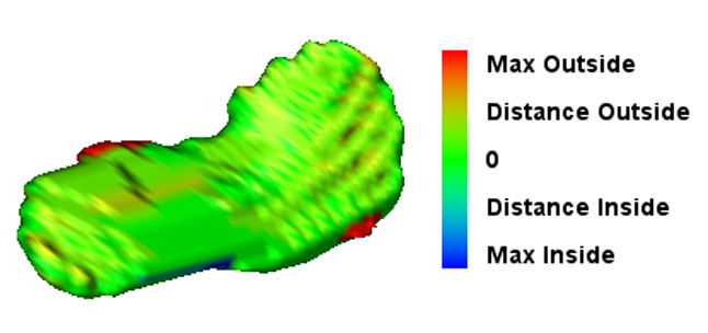
Degeneration of knee joint cartilage is an important and early indicator of osteoarthritis (OA) which is one of the major socio-economic burdens nowadays. Accurate quantification of the articular cartilage degeneration in an early stage using MR images is a promising approach in diagnosis and therapy for this disease. Particularly, volume and thickness measurement of cartilage tissue has been shown to deliver significant parameters in assessment of pathologies. Here, accurate computer-aided diagnosis tools could improve the clinical routine where image segmentation plays a crucial role. In order to overcome the time-consuming and tedious work of manual segmentation, one tries to automate the segmentation as much as possible. We focus on novel atlas-based segmentation methods for knee cartilage as well as improve today’s clinical routine of manual segmentation methods. In addition, we try to evaluate different methods for the assessment of parameters such as volume and thickness which could allow computer-aided diagnosis of knee cartilage pathologies in an early stage.Detailed Project Description
The early development of osteoarthritis (OA), a major socio-economic burden nowadays, is known to be related to the condition of the articular cartilage. For the development and refinement of therapies, noninvasive, accurate, and valid tools are needed to establish appropriate indications for new treatment options, to monitor the disease process and to control therapeutic efficacy.
Among the techniques for articular cartilage imaging, two modalities are the most promising: magnetic resonance imaging (MRI) and x-ray phase contrast imaging (PCI). Whereas in MRI image formation and acquisition is well-understood and research in cartilage examination concentrates on post-processing, in the field of PCI there is still need for adaption of existing or development of new acquisition techniques. In terms of evaluation of such methods for image acquistion and analysis, only histology is accepted to provide reliable gold standard data.
Magnetic Resonance Imaging

Main article: Semi-Automatic Patellar Cartilage Segmentation
MRI is a powerful tool for non-invasive examination of articular cartilage. In the analysis and interpretation of images, segmentation is a crucial step. This task still being too complex for fully automatic procedures, yet too tedious for completely manual accomplishment, only semi-automatic approaches permit contribution of radiologists’ expertise and acceptable expenditure of time. Such segmentations tools have been developed in recent years and are currently being evaluated.
Phase Contrast X-Ray Imaging
The emerging technique of phase contrast x-ray imaging is a promising modality for examination of soft tissue such as articular cartilage. The study of degenerative processes requires the development of volume reconstruction techniques for this modality, such as computerized tomography (CT) and tomosynthesis. The latter has been subject of a Diplomarbeit by Lorenz König.
Histology

Main article: Reconstruction and Registration of Histology and Phase Contrast Images for Clinical Validation of Imaging Modalities
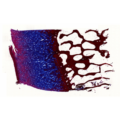
The gold standard in terms of evaluation of new imaging methods and modalities is histology. Currently, cross validation is performed by qualitative comparison of 3D datasets to 2D histology slices. For a more quantitative approach, we propose to improve the histology procedure and to develop methods towards a consistent reconstruction of 3D histology volumes.
Publications
Team
Contact Person(s)
|
Working Group
|
|
|
|
|
Alumni
|
Location
| Ludwig-Maximilians-Universität München Campus Grosshadern Marchioninistrae 15 81377 München Lab - Room: 4K U1 912 Tel.: +49 89 7095 4606 |
internal project page
Please contact Ben Glocker for available student projects within this research project.


