IDP: Development of an automatic multimodal in-situ visualisation tool
Advisor: Prof. Ekkehard EulerSupervision by: Joerg Traub, Tobias Sielhorst
Students:Latifa Omary, Philipp Stefan
Abstract

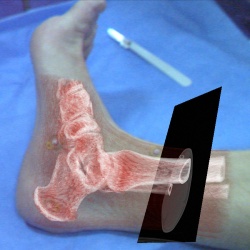
Figure 1: An in-situ visualisation of CT data showing a false colour volume rendering and an arbitrarily selected slice.
Introduction
For the treatment of spine fractures, that fulfil certain instability criteria
or suggest a kyphosis of more than 20°, the operative approach has become
widely accepted over the last two decades. While the type of access
however is still being dicussed controversely to date, it arises that the conventional
open access is connected with a considerably higher morbidity
than in minimally invasive procedures[4].
It was the reduction of access
morbidity without the need to accept diminished surgical effectiveness that
led to the development of endoscopic operations from representing exceptional
interventions to becoming standard procedures in spinal surgery[1].
In minimally invasive surgical approaches, the pre-operative planning phase
is of utmost importance, as the positions of the portals in relation to
one another and to the operating site significantly affect the entire course
of the operation. If the operating portal is not placed strictly above the injured
section, instruments can not be introduced in an orthograde position,
causing difficulties, that can lead to possible injury to major blood vessels
lying ventral to the spine or to the spinal cord, e.g. during the positioning
of intravertebral screws.
Therefore, it is inevitable for the surgeon to find - using the X-ray image
amplifier in combination with the preoperatively acquired CT data -
a proper projection of the target lesion, which is then drawn onto the skin
with a marker pen[1]. In order to place the portals using CT slice data
only as is, the surgeon has to look on a screen, posing the problem of mapping
the medical images onto the patient's body. Particularly the lack of
the third dimension makes this procedure very difficult. In consequence, it
demands considerable mental efforts and experience from the surgeon to
perform this task.
Scope of work
The goal of our project is to develop a visualisation tool, which provides the
surgeon with preoperatively acquired CT slices registered in 3D and augmented
directly onto the surgical object, using a video-see-through HMD.
This supplies the surgeon with a more intuitive view and a better spatial
feeling, hence giving higher precision during the placement of portals.
The only action required from the surgeon will be to attach markers to
the patient's skin in the vincinity of the surgical object and perform a CT
scan. Without any further interaction, he would then be able to instantly
see the result of the scan as an in-situ augmentation.

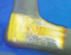
Figure 2: An in-situ visualisation of MRT data showing a false colour volume rendering.
Methods
The fiducial markers used are composed of standard CT skin markers,
which have a significant radiodensity, coated with IR retro-reflective material.
These properties make the markers detectable in both modalities,
the IR tracking and the CT scan. Furthermore each fiducial is constructed
in such a way that its centroid, in both modalities respectively, is located at
exactly the same spatial position, i.e. its center.
As a preparatory work, the centroids of the markers are automatically
calculated by applying common segmentation methods like for example region
growing algorithms to the CT data. Additionally, the infrared tracking
device will provide the up-to-date positions of the markers on the surgical
object. In order to find a transformation between the coordinate systems
defined by the markers in both modalities, the correspondences between
both point sets is automatically computed. Once the proper correspondences
are established, the rotation and translation between the two coordinate
systems can be estimated using 3D/3D registration methods[3].
This will enable the superimposition of the CT data visualisation on
the real environment. The visualisation methods used include a volume
rendering, enabling beside a real-time DRR from the user's point of view,
a false colour rendering using colour transfer functions. Another visualisation
method will provide the user with the possibility to view slices,
arbitrarily selected by a tracked "cutting plane".
Implementation
The implementation will be done using C++ and CAMPAR, a software toolkit developed at the chair of Computer Aided Medical Procedures. Our project involves the design of a new application within the framework of CAMPAR. Therefore already existent components can be used, but may be redefined. The visualisation will be based on the RAMP[2] hardware system and a commercial tracking system will be used to obtain the marker coordinates. All algorithms and hardware device interfaces that are needed to do tracking and accordingly segmentation and extraction of the marker positions from the CT data, as well as matching of corresponding points providing proper transformations, are to a certain degree already implemented. This reduces our effort considerably, enabling us to commit ourselves also to the demands on behalf of medical science.
Classification
The visualisation tool, called EVI - Easy Visualisation In-situ will be developed in the scope of an Interdisciplinary Project at the Chair for Computed Aided Medical Procedures at the Technical University of Munich in collaboration with the Chirurgische Klinik und Poliklinik - Innenstadt.
Remarks
The abovementioned imaging modality (CT) can substituted with several other medical imaging devices or methods, e.g. MRI, PET, Ultrasound etc. with reservations.
References[1] Buehren V. Beisse R., Potulski M. Endoscopic techniques for the management of spinal trauma. European Journal of Trauma, 27(6), 2001.
[2] Frank Sauer, Ali Khamene, and Sebastian Vogt. An augmented reality navigation system with a single-camera tracker: System design and needle biopsy phantom trial. In Proc. Int'l Conf. Medical Image Computing and Computer Assisted Intervention (MICCAI), 2002.
[3] Shinji Umeyama. Least-squares estimation of transformation parameters between two point patterns. IEEE Trans. Pattern Anal. Mach. Intell., 13(4):376–380, 1991.
[4] A.P. Verheyden, S. Katscher, O. Gonschorek, H. Lill, and C. Josten. Endoskopisch assistierte Rekonstruktion der thorakolumbalen Wirbelsäule in Bauchlage. Der Unfallchirurg, 105(10):873–880, 2002.
Milestones

Movie (AVI) of a live demo at ISMAR05
Useful Material & Links
Minutes
March 15, 2005
March 20, 2005
April 15, 2005
| ProjectForm | |
|---|---|
| Title: | Development of an automatic multimodal in-situ visualisation tool |
| Abstract: | For the treatment of spine fractures, that fulfil certain instability criteria, the minimally invasive approach has become widely accepted over the last decade. It was the reduction of access morbidity without the need to accept diminished surgical effectiveness that led to the development of endoscopic operations from representing exceptional interventions to becoming standard procedures in spinal surgery. In minimally invasive surgical approaches, the pre-operative planning phase is of utmost importance, as the positions of the portals in relation to one another and to the operating site significantly affect the entire course of the operation. The surgeon has to find - using the X-ray image amplifier in combination with the preoperatively acquired CT or MRI data - a proper projection of the target lesion onto the surgical area. This requires the surgeon to look on a screen, posing the problem of mentally mapping the medical images onto the patient's body. Particularly the lack of the third dimension makes this procedure very difficult. The goal of this project is the development of a visualisation tool, which provides the surgeon with preoperatively acquired imaging data registered in 3D and augmented directly onto the surgical object, using a video-see-through HMD. This would supply the surgeon with a more intuitive view and a better spatial feeling, hence yielding better results for the placement of portals. |
| Student: | Latifa Omary, Philipp Stefan |
| Director: | Prof. Ekkehard Euler |
| Supervisor: | Joerg Traub, Tobias Sielhorst |
| Type: | IDP |
| Status: | finished |
| Start: | 2005/04/15 |
| Finish: | 2005/12/31 |