ProjectZebrafish
Automatic thrombus segmentation in in vivo micoscopic video sequences under low contrast and highly dynamic conditionsIn academic collaboration with:Jovana Serbanovic-Canic, The Wellcome Trust Sanger Institute and the University of Cambridge; Dr. Ana Cvejic, The Wellcome Trust Sanger Institute, the University of Cambride; Dr. Willem Ouwehand, The Wellcome Trust Sanger Institute, the University of Cambride, and the National Health Service Blood and Transplant; Dr. Derek Stemple, The Wellcome Trust Sanger Institute; The Wellcome Trust Sanger Institute, Hixton, United Kingdom Departement of hematology, University of Cambridge, Cambridge, United Kingdom National Health Service Blood and Transplant, Cambridge, United Kingdom Contact Person(s): Nicolas Brieu |
Abstract
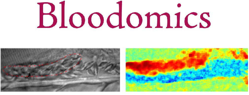
The Bloodomics EU project aims are to identify the genetic risks factors of coronary heart diseases.One of the most common methods is to study the thrombus formation in the dorsal aorta of mutant Zebrafish larvae. The developing thrombus is imaged in vivo through a microscope/camera setup. The derived time to attachment, growth speed, and time to occlusion permits the characterization of the thrombus formation.
However, this step presently remains manual. Our objective is to provide the geneticists in the Wellcome Trust Sanger Institute with an image processing tool to automatically detect and segment the growing thrombus. This will significantly speed up and improve the precision of this current analysis.
Publications
| 2012 | |
| N. Brieu, N. Navab, J. Serbanovic-Canic, W. Ouwehand
, D. Stemple, A. Cvejic, M. Groher
Image-based Characterization of Thrombus Formation in Time-lapse DIC Microscopy Medical Image Analysis 2012 (bib) |
|
| 2011 | |
| N. Brieu, M. Groher, J. Serbanovic-Canic, A. Cvejic, W. Ouwehand
, N. Navab
Joint Thrombus and Vessel Segmentation Using Dynamic Texture Likelihoods and Shape Prior Medical Image Computing and Computer-Assisted Intervention (MICCAI 2011), Toronto, Canada, September 2011 (bib) |
|
| 2010 | |
| N. Brieu, J. Serbanovic-Canic, A. Cvejic, D. Stemple, W. Ouwehand
, N. Navab, M. Groher
Thrombus Segmentation by Texture Dynamics from Microscopic Image Sequences SPIE Medical Imaging, 13-18 February 2010, San Diego, California, USA (bib) |
|
| N. Brieu, B. Glocker, N. Navab, M. Groher
MAP-MRF Optimal Partitioning for Dynamic Texture Segmentation of Thrombus in Time-Series Microscopic Images MICCAI 2010 Workshop on Spatio Temporal Image Analysis for Longitudinal and Time-Series Image Data (STIA'10), 24 September 2010, Beijing, China (bib) |
|
| 2009 | |
| N. Brieu, J. Serbanovic-Canic, A. Cvejic, D. Stemple, W. Ouwehand
, N. Navab, M. Groher
A dynamic texture approach to semi-automatic thrombosis segmentation in in-vivo microscopic video-sequences Workshop on Microscopic Image Analysis and Application in Biology, (MIAAB 2009), Bethesda, MD (US), 3-4 September 2009 (bib) |
|
Team
Contact Person(s)
|
Working Group
|
|
|
|
Location
| Technische Universität München Institut für Informatik / I16 Boltzmannstr. 3 85748 Garching bei München Tel.: +49 89 289-17058 Fax: +49 89 289-17059 |
internal project page
Please contact Nicolas Brieu for available student projects? within this research project.


