
Diploma Thesis: In-situ Visualization of Surgical Instruments in Medical Augmented Reality
Advisor: Nassir NavabSupervision by: Christoph Bichlmeier
Abstract
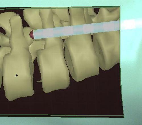
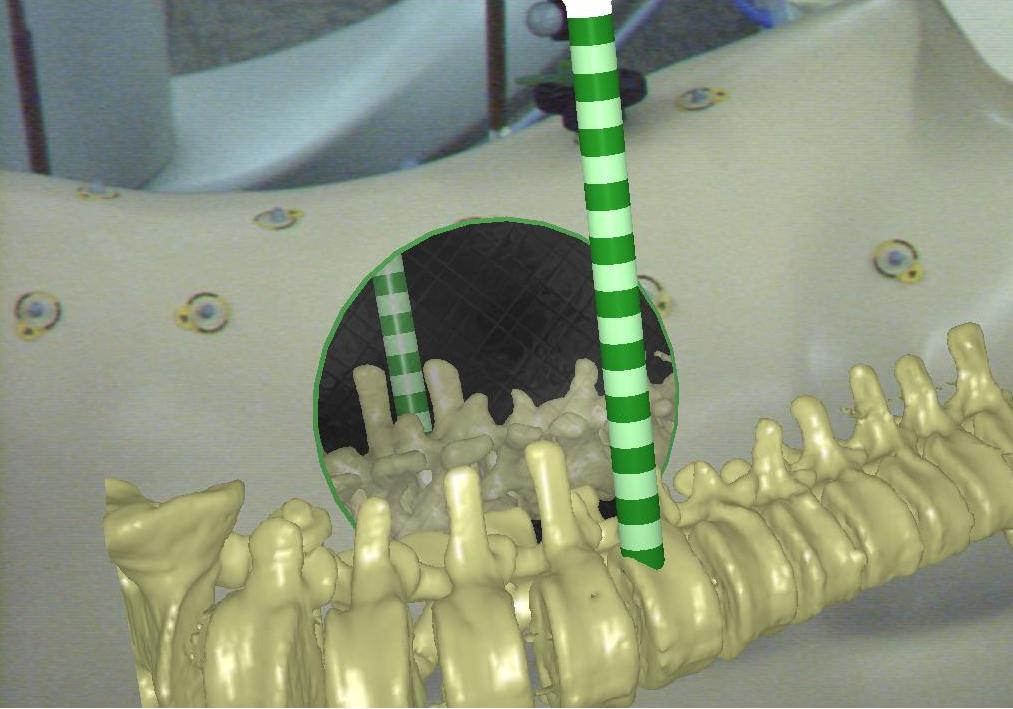
Medical augmented reality enables in-situ visualization of medical data. Tissue and bones e.g. the spinal column can be presented at its proper position at the patient using a stereoscopic head mounted display (HMD) or monitor based. Instruments, tracked by an external optical tracking system, can be augmented and guided inside the body of the patient where vision of the observer, in this case the surgeon, is restricted by the skin, blood and tissue. Beside navigated surgery such a system can be very helpful for teaching anatomy and for diagnosis. This project aims at the improvement of perceiving the relative and absolute position of tracked surgical instruments like a drill, resection tools, endoscope, etc inside the patient. After different experiments where in-situ visualization overlaid on phantoms was shown to the surgeons of our clinical partners, we identified the presentation of surgical instruments beside the presentation of medical data as one of the most important tasks to make this kind of data presentation acceptable. Here the surgeon is able interact with the medical data and knows about the position of the tip of the instrument when parts of the visualized medical data occlude the augmented instrument or the other way round. However, this is only one approach to provide visual perceptive cues due to interaction. You're task will be the integration of known interaction paradigms from computer graphics and create new ones aligned to the AR scenario. Presentation of medical data taken from CT or MRI scans can only be superimposed on real objects recorded by the cameras attached to the HMD. This means that virtual tissue and bones always occlude the real skin and seem to be located outside the human body. Your project will also aim at this problem. However, we have solutions for this task like a virtual window overlaid onto the skin, which can be integrated to your application. Finding the right visualization of surgical tools is very important to make Augmented Reality acceptable for different surgical tasks e.g. implantation of pedicle screws regarding spine surgery.Requirements
- Basic knowledge: C++, OpenGL, Computer Graphics (CG)
- Interest in medical technology, illustration
- Creativity and interest in design of 3D user interface
Your benefit
- Experience with different fields of research in computer science: Augmented Reality, Computer Graphics, Visualization, Software Engineering
- Knowledge about clinical/surgical workflow, medical technology
- Working within an interdisciplinary environment (You will work most of the time at Klinikum Innenstadt at Narvis).
Literature
| Students.ProjectForm | |
|---|---|
| Title: | In-situ Visualization of Surgical Instruments in Medical Augmented Reality |
| Abstract: | Medical augmented reality enables in-situ visualization of medical data. Tissue and bones e.g. the spinal column can be presented at its proper position at the patient using a stereoscopic head mounted display (HMD) or monitor based. Instruments, tracked by an external optical tracking system, can be augmented and guided inside the body of the patient where vision of the observer, in this case the surgeon, is restricted by the skin, blood and tissue. Beside navigated surgery such a system can be very helpful for teaching anatomy and for diagnosis. This project aims at the improvement of perceiving the relative and absolute position of tracked surgical instruments like a drill, resection tools, endoscope, etc inside the patient. After different experiments where in-situ visualization overlaid on phantoms was shown to the surgeons of our clinical partners, we identified the presentation of surgical instruments beside the presentation of medical data as one of the most important tasks to make this kind of data presentation acceptable. Here the surgeon is able interact with the medical data and knows about the position of the tip of the instrument when parts of the visualized medical data occlude the augmented instrument or the other way round. However, this is only one approach to provide visual perceptive cues due to interaction. You're task will be the integration of known interaction paradigms from computer graphics and create new ones aligned to the AR scenario. Presentation of medical data taken from CT or MRI scans can only be superimposed on real objects recorded by the cameras attached to the HMD. This means that virtual tissue and bones always occlude the real skin and seem to be located outside the human body. Your project will also aim at this problem. However, we have solutions for this task like a virtual window overlaid onto the skin, which can be integrated to your application. Finding the right visualization of surgical tools is very important to make Augmented Reality acceptable for different surgical tasks e.g. implantation of pedicle screws regarding spine surgery. |
| Student: | |
| Director: | Nassir Navab |
| Supervisor: | Christoph Bichlmeier |
| Type: | Diploma Thesis |
| Status: | open |
| Start: | Now |
| Finish: | Now + 6 month |