| 2020 |
|
A. Martin-Gomez
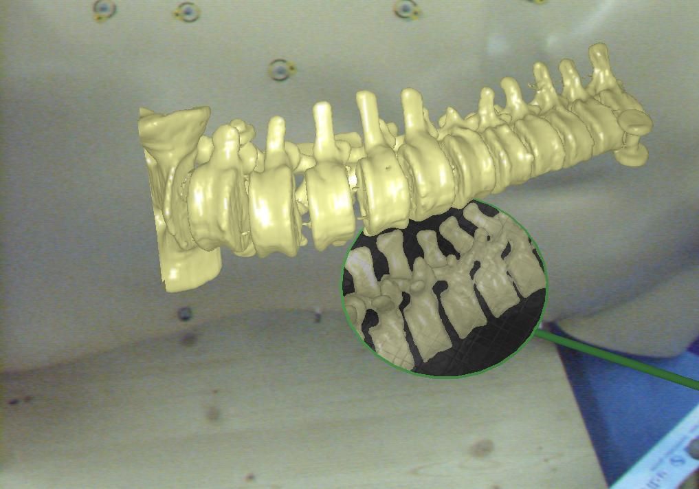
Evaluation of Different Visualization Techniques for Perception-Based Alignment in Medical AR
19th IEEE International Symposium on Mixed and Augmented Reality (ISMAR 2020)
(bib)
|
|
A. Martin-Gomez, J. Fotouhi, U. Eck, N. Navab
Gain A New Perspective: Towards Exploring Multi-View Alignment in Mixed Reality
19th IEEE International Symposium on Mixed and Augmented Reality (ISMAR 2020)
(bib)
|
|
A. Martin-Gomez, A. Winkler, K. Yu, T. Roth, U. Eck, N. Navab
Augmented Mirrors
19th IEEE International Symposium on Mixed and Augmented Reality (ISMAR 2020)
(bib)
|
|
A. Winkler, U. Eck, N. Navab
Spatially-Aware Displays for Computer Assisted Interventions
Proceedings of the 23rd International Conference on Medical Image Computing and Computer Assisted Interventions (MICCAI), Lima, October 2020
(bib)
|
|
A. Martin-Gomez, J. Fotouhi, N. Navab
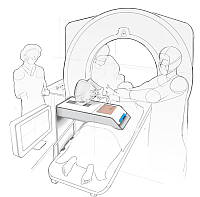
Towards Exploring the Benefits of Augmented Reality for Patient Support During Radiation Oncology Interventions
Computer Methods in Biomechanics and Biomedical Engineering: Imaging & Visualization
(bib)
|
|
A. Martin-Gomez, U. Eck, J. Fotouhi, N. Navab
Looking Also From Another Perspective: Exploring the Benefits of Alternative Views for Alignment Tasks
27th IEEE Virtual Reality Conference, Atlanta, Georgia, USA, Mar 2020
(bib)
|
| 2019 |
|
F. Bork, A. Lehner, D. Kugelmann, U. Eck, J. Waschke, N. Navab
VesARlius: An Augmented Reality System for Large-Group Co-Located Anatomy Learning
International Symposium on Mixed and Augmented Reality (ISMAR), 2019
(bib)
|
|
F. Bork, U. Eck, N. Navab
Birds vs. Fish: Visualizing Out-Of-View Objects in Augmented Reality using 3D Minimaps
International Symposium on Mixed and Augmented Reality (ISMAR), 2019
(bib)
|
|
A. Martin-Gomez, U. Eck, N. Navab
Visualization Techniques for Precise Alignment in VR. A Comparative Study
26th IEEE Virtual Reality Conference, Osaka, Japan, Mar 2019
(bib)
|
| 2018 |
|
F. Bork, A. Schnelzer, U. Eck, N. Navab
Towards Efficient Visual Guidance in Limited Field-of-View Head-Mounted Displays
IEEE Transactions on Visualization and Computer Graphics
(bib)
|
|
S. Matinfar, A. Nasseri, U. Eck, , A. Roodaki, Navid Navab, C. Lohmann, M. Maier, N. Navab
Surgical soundtracks: automatic acoustic augmentation of surgical procedures
International Journal of Computer Assisted Radiology and Surgery.
The final publication is available here
(bib)
|
|
U. Eck, T. Sielhorst
Display Technologies
Mixed and Augmented Reality in Medicine, CRC Press, 1st edition. 14pp, July 2018
(bib)
|
|
U. Eck, A. Winkler
Display-Technologien fuer Augmented Reality in der Medizin
Der Unfallchirurg, Springer International, Volume 121, Number 4 pp. 278--285, Feb 2018
(bib)
|
| 2017 |
|
F. Bork, R. Barmaki, U. Eck, K. Yu, C. Sandor, N. Navab
Empirical Study of Non-Reversing Magic Mirrors for Augmented Reality Anatomy Learning
International Symposium on Mixed and Augmented Reality (ISMAR), 2017
(bib)
|
|
S. Matinfar, A. Nasseri, U. Eck, A. Roodaki, Navid Navab, C. Lohmann, M. Maier, N. Navab
Surgical Soundtracks: Towards Automatic Musical Augmentation of Surgical Procedures
Proceedings of the 20th International Conference on Medical Image Computing and Computer Assisted Interventions (MICCAI), Quebec, Canada, September 2017. This work won the young scientist award at MICCAI 2017.
The final publication is available here
(bib)
|
|
S. Andress, U. Eck, C. Becker, A. Greiner, B. Rubenbauer, C. Linhart, S. Weidert
Automatic Surface Model Reconstruction to Enhance Treatment of Acetabular Fracture Surgery with 3D Printing
17TH ANNUAL MEETING OF THE INTERNATIONAL SOCIETY FOR COMPUTER ASSISTED ORTHOPAEDIC SURGERY, Aachen, Germany, pp. TBA, Jun 2017
(bib)
|
|
F. Bork, R. Barmaki, U. Eck, P. Fallavollita, B. Fuerst, N. Navab
Exploring Non-reversing Magic Mirrors for Screen-Based Augmented Reality Systems
24th IEEE Virtual Reality Conference, Los Angeles, CA, USA, pp. 373-374, Mar 2017
(bib)
|
| 2016 |
|
Alana Da Gama, Thiago Menezes Chaves, Lucas Silva Figueiredo, Adriana Baltar, Ma Meng, N. Navab, Veronica Teichrieb, P. Fallavollita
MirrARbilitation: A clinically-related gesture recognition interactive tool for an AR rehabilitation system
Computer Methods and Programs in Biomedicine
(bib)
|
|
Xiang Wang, S. Habert, C. Schulte-zu-Berge, P. Fallavollita, N. Navab
Inverse visualization concept for RGB-D augmented C-arms
Computers in Biology and Medicine
(bib)
|
|
Ma Meng, Philipp Jutzi, F. Bork, Ina Seelbach, A. von der Heide, N. Navab, P. Fallavollita
Interactive mixed reality for muscle structure and function learning
7th International Conference on Medical Imaging and Augmented Reality (to appear), Aug 2016
The first three authors contribute equally to this paper.
(bib)
|
|
U. Eck, P. Stefan, H. Laga, C. Sandor, P. Fallavollita, N. Navab
Exploring Visuo-Haptic Augmented Reality User Interfaces for Stereo-Tactic Neurosurgery Planning
7th International Conference on Medical Imaging and Augmented Reality, Bern, Switzerland, pp. 208-220, Aug 2016
(bib)
|
|
Ma Meng, P. Fallavollita, S. Habert, Simon Weider, N. Navab
Device and System Independent Personal Touchless User Interface for Operating Rooms
International Conference on Information Processing in Computer-Assisted Interventions (IPCAI), 2016
(bib)
|
|
P. Fallavollita, A. Brand, L. Wang, E. Euler, P. Thaller, N. Navab, S. Weidert
An augmented reality C-arm for intraoperative assessment of the mechanical axis - a preclinical study
International Journal of Computer Assisted Radiology and Surgery
(bib)
|
|
Xiang Wang, S. Habert, Ma Meng, C.-H. Huang, P. Fallavollita, N. Navab
Precise 3D/2D calibration between a RGB-D sensor and a C-arm fluoroscope
International Journal of Computer Assisted Radiology and Surgery
(bib)
|
|
Ma Meng, P. Fallavollita, Ina Seelbach, A. von der Heide, E. Euler, J. Waschke, N. Navab
Personalized augmented reality for anatomy education
Clinical Anatomy
(bib)
|
|
P. Fallavollita, L. Wang, S. Weidert, N. Navab
Augmented Reality in Orthopaedic Interventions and Education
Computational Radiology for Orthopaedic Interventions, Springer International Publishing 2016
(bib)
|
| 2015 |

|
M. Zweng, P. Fallavollita, S. Demirci, M. Kowarschik, N. Navab, D. Mateus
Automatic Guide-Wire Detection for Neurointerventions Using Low-Rank Sparse Matrix Decomposition and Denoising
MICCAI 2015 Workshop on Augmented Environments for Computer-Assisted Interventions
(bib)
|
|
Hagen Kaiser, P. Fallavollita, N. Navab
Real-Time Markerless Respiratory Motion Management Using Thermal Sensor Data
MICCAI 2015 Workshop on Augmented Environments for Computer-Assisted Interventions
(bib)
|
|
Hagen Kaiser, P. Fallavollita, N. Navab
'On the Fly' Reconstruction and Tracking System for Patient Setup in Radiation Therapy
MICCAI 2015 Workshop on Augmented Environments for Computer-Assisted Interventions
(bib)
|
|
R. Londei, M. Esposito, B. Diotte, S. Weidert, E. Euler, P. Thaller, N. Navab, P. Fallavollita
Intra-operative augmented reality in distal locking
The International Journal for Computer Assisted Radiology and Surgery (IJCARS)
(bib)
|
|
Xiang Wang, S. Habert, Ma Meng, C.-H. Huang, P. Fallavollita, N. Navab
RGB-D/C-arm Calibration and Application in Medical Augmented Reality
International Symposium on Mixed and Augmented Reality (ISMAR), 2015
(bib)
|
|
Ma Meng, , P. Fallavollita, N. Navab
Natural user interface for ambient objects
International Symposium on Mixed and Augmented Reality (ISMAR), 2015
(bib)
|
|
N. Leucht, S. Habert, P. Wucherer, S. Weidert, N. Navab, P. Fallavollita
Augmented Reality for Radiation Awareness
International Symposium on Mixed and Augmented Reality (ISMAR), 2015
(bib)
|
|
S. Habert, Ma Meng, W. Kehl, Xiang Wang, F. Tombari, P. Fallavollita, N. Navab
Augmenting mobile C-arm fluoroscopes via Stereo-RGBD sensors for multimodal visualization
International Symposium on Mixed and Augmented Reality (ISMAR), 2015
(bib)
|
|
S. Habert, J. Gardiazabal, P. Fallavollita, N. Navab
RGBDX: first design and experimental validation of a mirror-based RGBD Xray imaging system
International Symposium on Mixed and Augmented Reality (ISMAR), 2015
(bib)
|

|
F. Bork, B. Fuerst, A.K. Schneider, F. Pinto, C. Graumann, N. Navab
Auditory and Visio-Temporal Distance Coding for 3-Dimensional Perception in Medical Augmented Reality
International Symposium on Mixed and Augmented Reality (ISMAR), 2015
(bib)
|
|
C. Baur, F. Milletari, V. Belagiannis, N. Navab, P. Fallavollita
Automatic 3D reconstruction of electrophysiology catheters from two-view monoplane C-arm image sequences
The 6th International Conference on Information Processing in Computer-Assisted Interventions (IPCAI)
(bib)
|
|
M. Weigl, P. Stefan, K. Abhari, P. Wucherer, P. Fallavollita, M. Lazarovici, S. Weidert, K. Catchpole
Intra-operative disruptions, surgeon mental workload, and technical performance in a full-scale simulated procedure
Surgical Endoscopy
(bib)
|
|
N. Navab, P. Fallavollita, S. Weidert, E. Euler
CAMP@NARVIS: A Real-World AR/VR Research Laboratory
IEEE Virtual Reality
(bib)
|
|
R. Eagleson, P. Wucherer, P. Stefan, Y. Duschko, S. deRibaupierre, C. Vollmar, P. Fallavollita, N. Navab
Collaborative Table-Top VR Display for Neurosurgical Planning
IEEE Virtual Reality
(bib)
|
|
P. Wucherer, P. Stefan, K. Abhari, P. Fallavollita, M. Weigl, M. Lazarovici, A. Winkler, S. Weidert, T. Peters, S. deRibaupierre, R. Eagleson, N. Navab
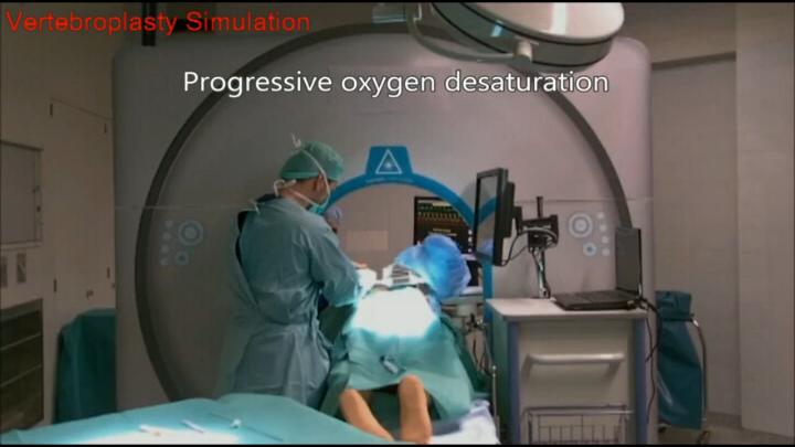
Vertebroplasty Performance on Simulator for 19 Surgeons Using Hierarchical Task Analysis
IEEE Transactions on Medical Imaging
(bib)
|
| 2014 |
|
O. Pauly, B. Diotte, P. Fallavollita, S. Weidert, E. Euler, N. Navab
Machine Learning-based Augmented Reality for Improved Surgical Scene Understanding.
Computerized Medical Imaging and Graphics
(bib)
|
|
O. Pauly, B. Diotte, S. Habert, S. Weidert, E. Euler, P. Fallavollita, N. Navab
Visualization inside the operating room: 'Learning' what the surgeon wants to see
The 5th International Conference on Information Processing in Computer-Assisted Interventions (IPCAI)
(bib)
|
|
R. Londei, M. Esposito, B. Diotte, S. Weidert, E. Euler, P. Thaller, N. Navab, P. Fallavollita
The Augmented Circles: A Video-Guided Solution for the Down-the-Beam Positioning of IM Nail Holes
The 5th International Conference on Information Processing in Computer-Assisted Interventions (IPCAI)
(bib)
|
|
B. Diotte, P. Fallavollita, L. Wang, S. Weidert, E. Euler, P. Thaller, N. Navab
Multi-modal intra-operative navigation during distal locking of intramedullary nails
IEEE Transactions on Medical Imaging
(bib)
|


|
F. Milletari, V. Belagiannis, N. Navab, P. Fallavollita
Fully automatic catheter localization in C-arm images using l1- Sparse Coding
Proceedings of the 17th International Conference on Medical Image Computing and Computer Assisted Interventions (MICCAI), Boston, September 2014
(bib)
|
|
P. Fallavollita, A. Winkler, S. Habert, P. Wucherer, P. Stefan, Riad Mansour, R. Ghotbi, N. Navab
'Desired-View' controlled positioning of angiographic C-arms
Proceedings of the 17th International Conference on Medical Image Computing and Computer Assisted Interventions (MICCAI), Boston, September 2014
(bib)
|
|
Xiang Wang, S. Demirci, C. Schulte-zu-Berge, P. Fallavollita, N. Navab
Improved Interventional X-ray Appearance
International Symposium on Mixed and Augmented Reality (ISMAR), 2014
(bib)
|
|
Alana Da Gama, P. Fallavollita, Veronica Teichrieb, N. Navab
Motor rehabilitation using Kinect: a systematic review
Games for Health Journal
(bib)
|
|
Jian Wang, L. Wang, Matthias Kreiser, N. Navab, P. Fallavollita
Augmented Depth Perception Visualization in 2D/3D Image Fusion
Computerized Medical Imaging and Graphics
(bib)
|
|
X. Chen, Hemal Naik, L. Wang, N. Navab, P. Fallavollita
Video Guided Calibration of an Augmented Reality mobile C-arm
International Journal of Computer Assisted Radiology and Surgery (IJCARS)
(bib)
|
|
P. Fallavollita
Detection, Tracking and Related Costs of Ablation Catheters in the Treatment of Cardiac Arrhythmias
Cardiac Arrhythmias - Mechanisms, Pathophysiology, and Treatment, Publisher: INTECH, Editor: Wilbert S. Aronow
(bib)
|
|
P. Wucherer, P. Stefan, S. Weidert, P. Fallavollita, N. Navab
Task and Crisis Analysis during Surgical Training
International Journal for Computer Assisted Radiology and Surgery (IJCARS)
(bib)
|
|
P. Stefan, P. Wucherer, Y. Oyamada, Ma Meng, A. Schoch, , , , , M. Weigl, , P. Fallavollita, H. Saito, N. Navab
An AR Edutainment System Supporting Bone Anatomy Learning
IEEE Virtual Reality
(bib)
|
|
C. Amat di San Filippo, G. Fichtinger, William Morris, Tim Salcudean, E. Dehghan, P. Fallavollita
Intraoperative Segmentation of Iodine and Palladium Radioactive Sources in C-arm images
International Journal for Computer Assisted Radiology and Surgery (IJCARS)
(bib)
|
| 2013 |
|
S. Weidert, P. Wucherer, P. Stefan, S.P. Baierl, M. Weigl, M. Lazarovici, P. Fallavollita, N. Navab
A Vertebroplasty simulation environment for medical education.
Bone Joint J, 2013
(bib)
|
|
P. Wucherer, P. Stefan, S. Weidert, P. Fallavollita, N. Navab
Development and procedural evaluation of immersive medical simulation environments
The 4th International Conference on Information Processing in Computer-Assisted Interventions (IPCAI), Heidelberg, Germany, June 26, 2013
(bib)
|
|
P. Thaller, B. Diotte, P. Fallavollita, L. Wang, S. Weidert, E. Euler, W. Mutschler, N. Navab
Video-assisted interlocking of intramedullary nails
Injury, 2013
(bib)
|
|
F. Milletari, N. Navab, P. Fallavollita
Automatic detection of multiple and overlapping EP catheters in fluoroscopic sequences
Proceedings of the 16th International Conference on Medical Image Computing and Computer Assisted Interventions (MICCAI), Nagoya, Japan, September 2013
(bib)
|
|
P. Thaller, B. Diotte, P. Fallavollita, L. Wang, S. Weidert, W. Mutschler, N. Navab, E. Euler
Video-navigated targeting unit for interlocking of intramedullary nails – experimental study on a new technique.
Kongress der Gesellschaft für Extremitätenverlängerung und Rekonstruktion, 2013
(bib)
|
|
C. Amat di San Filippo, G. Fichtinger, William Morris, Tim Salcudean, E. Dehghan, P. Fallavollita
Declustering n-connected components: an example case for the segmentation of iodine implants in C-arm images
4th International Conference on Information Processing in Computer-Assisted Interventions (IPCAI), Heidelberg, Germany, June 26, 2013
(bib)
|
|
S. Weidert, P. Wucherer, P. Stefan, P. Fallavollita, E. Euler, N. Navab
Vertebroplasty medical simulation learning environment
International Society for Computer Assisted Orthopaedic Surgery, Orlando, June 2013
(bib)
|
|
S. Pati, O. Erat, L. Wang, S. Weidert, E. Euler, N. Navab, P. Fallavollita
Accurate pose estimation using a single marker camera calibration system
SPIE Medical Imaging, Orlando, Florida, USA, February 2013
(bib)
|
|
O. Erat, O. Pauly, S. Weidert, P. Thaller, E. Euler, W. Mutschler, N. Navab, P. Fallavollita
How a Surgeon becomes Superman by visualization of intelligently fused multi-modalities
SPIE Medical Imaging, Orlando, Florida, USA, February 2013
(bib)
|
|
Ma Meng, P. Fallavollita, T. Blum, U. Eck, C. Sandor, S. Weidert, J. Waschke, N. Navab
Kinect for Interactive AR Anatomy Learning
The 12th IEEE and ACM International Symposium on Mixed and Augmented Reality (ISMAR), Adelaide, Australia, Oct. 1 - 4, 2013.
(bib)
|
| 2012 |
|
A. Ahmadi, T. Klein, A. Plate, K. Bötzel, N. Navab
Rigid US-MRI Registration Through Segmentation of Equivalent Anatomic Structures - A feasibility study using 3D transcranial ultrasound of the midbrain.
Workshop Bildverarbeitung fuer die Medizin, Berlin (GER), March 18-20, 2012
(bib)
|
|
Y. Oyamada, P. Fallavollita, N. Navab
Single Camera Calibration using partially visible calibration objects based on Random Dots Marker Tracking Algorithm
The IEEE and ACM International Symposium on Mixed and Augmented Reality (ISMAR), Workshop on Tracking Methods and Applications, 2012
(bib)
|
|
Jian Wang, P. Fallavollita, L. Wang, Matthias Kreiser, N. Navab
Augmented Reality during Angiography: Integration of a Virtual Mirror for Improved 2D/3D Visualization
The 11th IEEE and ACM International Symposium on Mixed and Augmented Reality (ISMAR), Atlanta, USA, Nov. 5 - 8, 2012.
(bib)
|
|
O. Pauly, A. Katouzian, A. Eslami, P. Fallavollita, N. Navab
Supervised Classification for Customized Intraoperative Augmented Reality Visualization
The 11th IEEE and ACM International Symposium on Mixed and Augmented Reality (ISMAR), Atlanta, USA, Nov. 5 - 8, 2012.
(bib)
|

|
T. Blum, R. Stauder, E. Euler, N. Navab
Superman-like X-ray Vision: Towards Brain-Computer Interfaces for Medical Augmented Reality
The 11th IEEE and ACM International Symposium on Mixed and Augmented Reality (ISMAR), Atlanta, USA, Nov. 5 - 8, 2012. The original publication is available online at ieee.org.
(bib)
|
|
A. Aichert, M. Wieczorek, Jian Wang, L. Wang, Matthias Kreiser, P. Fallavollita, N. Navab
The Colored X-rays
MICCAI 2012 Workshop on Augmented Environments for Computer-Assisted Interventions
(bib)
|
|
L. Wang, P. Fallavollita, A. Brand, O. Erat, S. Weidert, P. Thaller, E. Euler, N. Navab
Intra-op measurement of the mechanical axis deviation: an evaluation study on 19 human cadaver legs
Proceedings of the 15th International Conference on Medical Image Computing and Computer Assisted Interventions (MICCAI), Nice, France, October 2012
(bib)
|


|
B. Diotte, P. Fallavollita, L. Wang, S. Weidert, P. Thaller, E. Euler, N. Navab
Radiation-free drill guidance in interlocking of intramedullary nails
Proceedings of the 15th International Conference on Medical Image Computing and Computer Assisted Interventions (MICCAI), Nice, France, October 2012
(bib)
|
|
A. Plate, A. Ahmadi, O. Pauly, T. Klein, N. Navab, K. Bötzel
3D Sonographic Examination of the Midbrain for Computer-Aided Diagnosis of Movement Disorders
To appear: Ultrasound in Medicine and Biology, ELSEVIER, Christy K. Holland (Editor), accepted for publication July 21 2012
(bib)
|
|
E. Dehghan, J. Lee, P. Fallavollita, N. Kuo, A. Deguet, Yi Le, C. Burdette, D. Song, J. Prince, G. Fichtinger
Ultrasound-Fluoroscopy Registration for Prostate Brachytherapy Dosimetry
Medical Image Analysis, 2012
(bib)
|
|
A. Brand, L. Wang, P. Fallavollita, Philip Sandner, N. Navab, P. Thaller, E. Euler, S. Weidert
Intraoperative measurement of mechanical axis alignment by automatic image stitching: a human cadaver study
International Society for Computer Assisted Orthopaedic Surgery (CAOS), South Korea, June 2012
(bib)
|
|
S. Weidert, L. Wang, A. von der Heide, N. Navab, E. Euler
Intraoperative augmented-reality-visualisierung: Aktueller stand der entwicklung und erste erfahrungen mit dem camc
Unfallchirurg, 2012 (Accepted for publication)
(bib)
|
|
P. Fallavollita
The Future of Cardiac Mapping
Cardiac Arrhythmias - New Considerations, Publisher: INTECH, Editor: Francisco R. Breijo-Marquez
(bib)
|
|
L. Wang, P. Fallavollita, R. Zou, X. Chen, S. Weidert, N. Navab
Closed-form inverse kinematics for interventional C-arm X-ray imaging with six degrees of freedom: modeling and application
IEEE Transactions on Medical Imaging
(bib)
|
|
C. Wachinger, M. Yigitsoy, E. Rijkhorst, N. Navab
Manifold Learning for Image-Based Breathing Gating in Ultrasound and MRI
Medical Image Analysis, Volume 16, Issue 4, May 2012, Pages 806–818.
(bib)
|
| 2011 |


|
M. Wieczorek, A. Aichert, P. Fallavollita, O. Kutter, A. Ahmadi, L. Wang, N. Navab
Interactive 3D visualization of a single-view X-Ray image
Proceedings of Medical Image Computing and Computer-Assisted Intervention (MICCAI 2011), Toronto, Canada, September 2011.
(bib)
|
|
S. Weidert, L. Wang, P. Thaller, J. Landes, A. Brand, N. Navab, E. Euler
X-ray Stitching for Intra-operative Mechanical Axis Determination of the Lower Extremity
the 11th Annual Meeting of the International Society for Computer Assisted Orthopaedic Surgery, London, UK, June 15-19 2011
(bib)
|
|
L. Wang, R. Zou, S. Weidert, J. Landes, E. Euler, D. Burschka, N. Navab
Closed-form inverse kinematics for intra-operative mobile C-arm positioning with six degrees of freedom
SPIE Medical Imaging, Lake Buena Vista (Orlando), Florida, USA, February 2011
(bib)
|
| 2010 |
|
C. Bichlmeier
Immersive, Interactive and Contextual In-Situ Visualization for Medical Applications
Dissertation an der Fakultät für Informatik, Technische Universität München, 2010 ( online version available here)
(bib)
|
|
L. Wang, M. Springer, H. Heibel, N. Navab
Floyd-Warshall All-Pair Shortest Path for Accurate Multi-Marker Calibration
The 9th IEEE and ACM International Symposium on Mixed and Augmented Reality, Seoul, Korea, Oct. 13 - 16, 2010.
(bib)
|


|
T. Blum, M. Wieczorek, A. Aichert, R. Tibrewal, N. Navab
The effect of out-of-focus blur on visual discomfort when using stereo displays
The 9th IEEE and ACM International Symposium on Mixed and Augmented Reality, Seoul, Korea, Oct. 13 - 16, 2010. The original publication is available online at ieee.org.
(bib)
|

|
C. Bichlmeier, E. Euler, T. Blum, N. Navab
Evaluation of the Virtual Mirror as a Navigational Aid for Augmented Reality Driven Minimally Invasive Procedures
The 9th IEEE and ACM International Symposium on Mixed and Augmented Reality, Seoul, Korea, Oct. 13 - 16, 2010. The original publication is available online at ieee.org.
(bib)
|
|
L. Wang, R. Zou, S. Weidert, J. Landes, E. Euler, D. Burschka, N. Navab
Modeling Kinematics of Mobile C-arm and Operating Table as an Integrated Six Degrees of Freedom Imaging System
The 5th International Workshop on Medical Imaging and Augmented Reality, MIAR 2010, Beijing, China, September 19-20, 2010
(bib)
|
|
A. Ahmadi, F. Pisana, E. DeMomi?, N. Navab, G. Ferrigno
User friendly graphical user interface for workflow management during navigated robotic-assisted keyhole neurosurgery
Computer Assisted Radiology (CARS), 24th International Congress and Exhibition, Geneva, CH, June 2010
(bib)
|
|
L. Wang, J. Traub, S. Weidert, S.M. Heining, E. Euler, N. Navab
Parallax-Free Intra-Operative X-ray Image Stitching
the MICCAI 2009 special issue of the Journal Medical Image Analysis
(bib)
|

|
L. Wang, J. Landes, S. Weidert, T. Blum, A. von der Heide, E. Euler, N. Navab
First Animal Cadaver Study for Interlocking of Intramedullary Nails under Camera Augmented Mobile C-arm A Surgical Workflow Based Preclinical Evaluation
the 1st International Conference on Information Processing in Computer-Assisted Interventions (IPCAI), Switzerland, June 23 2010. The original publication is available online at www.springerlink.com.
(bib)
|
|
S. Weidert, L. Wang, J. Landes, A. von der Heide, N. Navab, E. Euler
First Surgical Procedures under Camera-Augmented Mobile C-arm (CamC) guidance
The 3rd Hamlyn Symposium for Medical Robotics ,London, UK, May 25 2010
(bib)
|
|
A. Plate, A. Ahmadi, T. Klein, N. Navab, J. Weiße, J. Mehrkens, K. Bötzel
Towards a More Objective Visualization of the Midbrain and its Surroundings Using 3D Transcranial Ultrasound
54. Jahrestagung der Deutschen Gesellschaft für Klinische Neurophysiologie und Funktionelle Bildgebung (DGKN), Halle, GER, March 2010
(bib)
|

|
P. Wucherer, C. Bichlmeier, M. Eder, L. Kovacs, N. Navab
Multimodal Medical Consultation for Improved Patient Education
Proceedings of Bildverarbeitung fuer die Medizin (BVM 2010), Aachen, Germany, March 2010
(bib)
|

|
M. Wieczorek, A. Aichert, O. Kutter, C. Bichlmeier, J. Landes, S.M. Heining, E. Euler, N. Navab
GPU-accelerated Rendering for Medical Augmented Reality in Minimally-Invasive Procedures
Proceedings of Bildverarbeitung fuer die Medizin (BVM 2010), Aachen, Germany, March 14-16 2010
(bib)
|

|
P. Dressel, L. Wang, O. Kutter, J. Traub, S.M. Heining, N. Navab
Intraoperative positioning of mobile C-arms using artificial fluoroscopy
SPIE Medical Imaging, San Diego, California, USA, February 2010
(bib)
|
| 2009 |

|
C. Bichlmeier, S.M. Heining, L. Omary, P. Stefan, B. Ockert, E. Euler, N. Navab
MeTaTop: A Multi-Sensory and Multi-User Interface for Collaborative Analysis of Medical Imaging Data
Interactive Demo (ITS 2009), Banff, Canada, November 2009
(bib)
|

|
C. Bichlmeier, S. Holdstock, S.M. Heining, S. Weidert, E. Euler, O. Kutter, N. Navab
Contextual In-Situ Visualization for Port Placement in Keyhole Surgery: Evaluation of Three Target Applications by Two Surgeons and Eighteen Medical Trainees
The 8th IEEE and ACM International Symposium on Mixed and Augmented Reality, Orlando, US, Oct. 19 - 22, 2009.
(bib)
|

|
C. Bichlmeier, M. Kipot, S. Holdstock, S.M. Heining, E. Euler, N. Navab
A Practical Approach for Intraoperative Contextual In-Situ Visualization
International Workshop on Augmented environments for Medical Imaging including Augmented Reality in Computer-aided Surgery (AMI-ARCS 2009), London, UK, September 2009
(bib)
|

|
C. Bichlmeier, S.M. Heining, M. Feuerstein, N. Navab
The Virtual Mirror: A New Interaction Paradigm for Augmented Reality Environments
IEEE Trans. Med. Imag., vol. 28, no. 9, pp. 1498-1510, September 2009
(bib)
|
|
L. Wang, J. Traub, S. Weidert, S.M. Heining, E. Euler, N. Navab
Parallax-free Long Bone X-ray Image Stitching
Medical Image Computing and Computer-Assisted Intervention (MICCAI), London, UK, September 20-24 2009
(bib)
|
|
A. Ahmadi, T. Klein, N. Navab
Advanced Planning and Ultrasound Guidance for Keyhole Neurosurgery in ROBOCAST
Russian Bavarian Conference (RBC), Munich, GER, July 2009
(bib)
|
|
A. Ahmadi, T. Klein, N. Navab, R. Roth, R.R. Shamir, L. Joskowicz, E. DeMomi?, G. Ferrigno, L. Antiga, R.I. Foroni
Advanced Planning and Intra-operative Validation for Robot-Assisted Keyhole Neurosurgery In ROBOCAST
International Conference on Advanced Robotics (ICAR), Munich, GER, June 2009
(bib)
|

|
B. Ockert, C. Bichlmeier, S.M. Heining, O. Kutter, N. Navab, E. Euler
Development of an Augmented Reality (AR) training environment for orthopedic surgery procedures
Proceedings of The 9th Computer Assisted Orthopaedic Surgery (CAOS 2009), Boston, USA, June, 2009
(bib)
|

|
N. Navab, S.M. Heining, J. Traub
Camera Augmented Mobile C-arm (CAMC): Calibration, Accuracy Study and Clinical Applications
IEEE Transactions Medical Imaging, 29 (7), 1412-1423
(bib)
|
|
L. Wang, J. Traub, S.M. Heining, S. Benhimane, R. Graumann, E. Euler, N. Navab
Long Bone X-ray Image Stitching using C-arm Motion Estimation
Proceedings of Bildverarbeitung fuer die Medizin (BVM 2009), Heidelberg, Germany, March 22-24 2009
(bib)
|
|
L. Wang, S. Weidert, J. Traub, S.M. Heining, C. Riquarts, E. Euler, N. Navab
Camera Augmented Mobile C-arm: Towards Real Patient Study
Proceedings of Bildverarbeitung fuer die Medizin (BVM 2009), Heidelberg, Germany, March 22-24 2009
(bib)
|
| 2008 |
|
C. Bichlmeier, B. Ockert, S.M. Heining, A. Ahmadi, N. Navab
Stepping into the Operating Theater: ARAV - Augmented Reality Aided Vertebroplasty
The 7th IEEE and ACM International Symposium on Mixed and Augmented Reality, Cambridge, UK, Sept. 15 - 18, 2008.
(bib)
|
|
J. Traub
New Concepts for Design and Workflow Driven Evaluation of Computer Assisted Surgery Solutions
Dissertation an der Fakultät für Informatik, Technische Universität München, 2008 ( online version available here)
(bib)
|


|
O. Kutter, A. Aichert, C. Bichlmeier, J. Traub, S.M. Heining, B. Ockert, E. Euler, N. Navab
Real-time Volume Rendering for High Quality Visualization in Augmented Reality
International Workshop on Augmented environments for Medical Imaging including Augmented Reality in Computer-aided Surgery (AMI-ARCS 2008), USA, New York, September 2008
(bib)
|

|
C. Bichlmeier, B. Ockert, O. Kutter, M. Rustaee, S.M. Heining, N. Navab
The Visible Korean Human Phantom: Realistic Test & Development Environments for Medical Augmented Reality
International Workshop on Augmented environments for Medical Imaging including Augmented Reality in Computer-aided Surgery (AMI-ARCS 2008), USA, New York, September 2008
(bib)
|
|
J. Traub, A. Ahmadi, N. Padoy, L. Wang, S.M. Heining, E. Euler, P. Jannin, N. Navab
Workflow Based Assessment of the Camera Augmented Mobile C-arm System
International Workshop on Augmented Reality environments for Medical Imaging and Computer-aided Surgery (AMI-ARCS 2008), New York, NY, USA, September 2008
(bib)
|
|
L. Wang, J. Traub, S.M. Heining, S. Benhimane, R. Graumann, E. Euler, N. Navab
Long Bone X-ray Image Stitching Using Camera Augmented Mobile C-arm
Medical Image Computing and Computer-Assisted Intervention, MICCAI, 2008, New York, USA, September 6-10 2008
(bib)
|

|
F. Wimmer, C. Bichlmeier, S.M. Heining, N. Navab
Creating a Vision Channel for Observing Deep-Seated Anatomy in Medical Augmented Reality
Proceedings of Bildverarbeitung fuer die Medizin (BVM 2008), Munich, Germany, April 2008
(bib)
|
|
J. Traub, S.M. Heining, E. Euler, N. Navab
Two camera augmented mobile C-arm – System setup and first experiments
Proceedings of The 8th Computer Assisted Orthopaedic Surgery (CAOS 2008), Hong Kong, China, June, 2008
(bib)
|
|
S.M. Heining, C. Bichlmeier, E. Euler, N. Navab
Smart Device: Virtually Extended Surgical Drill
Proceedings of The 8th Computer Assisted Orthopaedic Surgery (CAOS 2008), Hong Kong, China, June, 2008
(bib)
|
|
T. Sielhorst
New Methods for Medical Augmented Reality
Dissertation an der Fakultät für Informatik, Technische Universität München, 2008 ( online version available here)
(bib)
|
| 2007 |


|
C. Bichlmeier, S.M. Heining, M. Rustaee, N. Navab
Laparoscopic Virtual Mirror for Understanding Vessel Structure: Evaluation Study by Twelve Surgeons
The Sixth IEEE and ACM International Symposium on Mixed and Augmented Reality, Nara, Japan, Nov. 13 - 16, 2007.
(bib)
|


|
C. Bichlmeier, F. Wimmer, S.M. Heining, N. Navab
Contextual Anatomic Mimesis: Hybrid In-Situ Visualization Method for Improving Multi-Sensory Depth Perception in Medical Augmented Reality
The Sixth IEEE and ACM International Symposium on Mixed and Augmented Reality, Nara, Japan, Nov. 13 - 16, 2007.
(bib)
|


|
T. Sielhorst, Wu Sa, A. Khamene, F. Sauer, N. Navab
Measurement of absolute latency for video see through augmented reality
Sixth IEEE and ACM International Symposium on Mixed and Augmented Reality (ISMAR'07), Nara, Japan, November 2007
(bib)
|


|
T. Sielhorst, M. Bauer, O. Wenisch, G. Klinker, N. Navab
Online Estimation of the Target Registration Error for n-ocular Optical Tracking Systems
to appear Proceedings of Medical Image Computing and Computer-Assisted Intervention (MICCAI 2007), Brisbane, Australia, October 2007, pp. 652-659.
(bib)
|

|
P. Stefan, J. Traub, S.M. Heining, C. Riquarts, T. Sielhorst, E. Euler, N. Navab
Hybrid navigation interface: a comparative study
Proceedings of Bildverarbeitung fuer die Medizin (BVM 2007), Munich, Germany, March 2007, pp. 81-86
(bib)
|

|
C. Bichlmeier, T. Sielhorst, S.M. Heining, N. Navab
Improving Depth Perception in Medical AR: A Virtual Vision Panel to the Inside of the Patient
Proceedings of Bildverarbeitung fuer die Medizin (BVM 2007), Munich, Germany, March 2007
(bib)
|
| 2006 |

|
C. Bichlmeier, N. Navab
Virtual Window for Improved Depth Perception in Medical AR
International Workshop on Augmented Reality environments for Medical Imaging and Computer-aided Surgery (AMI-ARCS 2006), Copenhagen, Denmark, October 2006
(bib)
|

|
J. Traub, P. Stefan, S.M. Heining, T. Sielhorst, C. Riquarts, E. Euler, N. Navab
Hybrid navigation interface for orthopedic and trauma surgery
Proceedings of Medical Image Computing and Computer-Assisted Intervention (MICCAI 2006), Copenhagen, Denmark, October 2006, pp. 373-380
(bib)
|


|
T. Sielhorst, C. Bichlmeier, S.M. Heining, N. Navab
Depth perception a major issue in medical AR: Evaluation study by twenty surgeons
Proceedings of Medical Image Computing and Computer-Assisted Intervention (MICCAI 2006), Copenhagen, Denmark, October 2006, pp. 364-372
The original publication is available online at www.springerlink.com
(bib)
|

|
J. Traub, P. Stefan, S.M. Heining, T. Sielhorst, C. Riquarts, E. Euler, N. Navab
Towards a Hybrid Navigation Interface: Comparison of a Slice Based Navigation System with In-situ Visualization
Proceedings of International Workshop on Medical Imaging and Augmented Reality (MIAR 2006), Shanghai, China, August, 2006, pp.179-186
(bib)
|

|
S.M. Heining, P. Stefan, L. Omary, S. Wiesner, T. Sielhorst, N. Navab, F. Sauer, E. Euler, W. Mutschler, J. Traub
Evaluation of an in-situ visualization system for navigated trauma surgery
Journal of Biomechanics 2006; Vol. 39 Suppl. 1, page 209
(bib)
|

|
J. Traub, P. Stefan, S.M. Heining, T. Sielhorst, C. Riquarts, E. Euler, N. Navab
Stereoscopic augmented reality navigation for trauma surgery: cadaver experiment and usability study
International Journal of Computer Assisted Radiology and Surgery, 2006; Vol. 1 Suppl. 1, page 30 - 31. The original publication is available online at www.springerlink.com
(bib)
|

|
T. Sielhorst, M. Feuerstein, J. Traub, O. Kutter, N. Navab
CAMPAR: A software framework guaranteeing quality for medical augmented reality
International Journal of Computer Assisted Radiology and Surgery, 2006; Vol. 1 Suppl. 1, page 29 - 30.
The original publication is available online at www.springerlink.com
(bib)
|
|
S.M. Heining, P. Stefan, F. Sauer, E. Euler, N. Navab, J. Traub
Evaluation of an in-situ visualization system for navigated trauma surgery
Proceedings of The 6th Computer Assisted Orthopaedic Surgery (CAOS 2006), Montreal, Canada, June, 2006
(bib)
|

