Andrei Chekkoury
|
M.Sc. Andrei Chekkoury
|
Active Research Projects
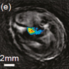
Methods for 3D Multispectral Tomography, Institute for Biological and Medical Imaging, prof. Ntziachristos |
|
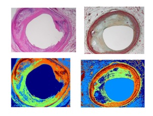
Automatic Labeling of Atherosclerotic Tissues in Histology ImagesAbstract: In this project, the goal is to automatically label atherosclerotic tissues in histology images that are often used to study and understand progression of atherosclerosis deploying different intravascular imaging modalities such IVUS, OCT, etc. The results can then be used to manual labeling of tissue for feature extraction and construction of reliable training dataset. It can further be used for systematic quantification of atherosclerotic tissue characterization algorithms. |
Previous Research Projects
|
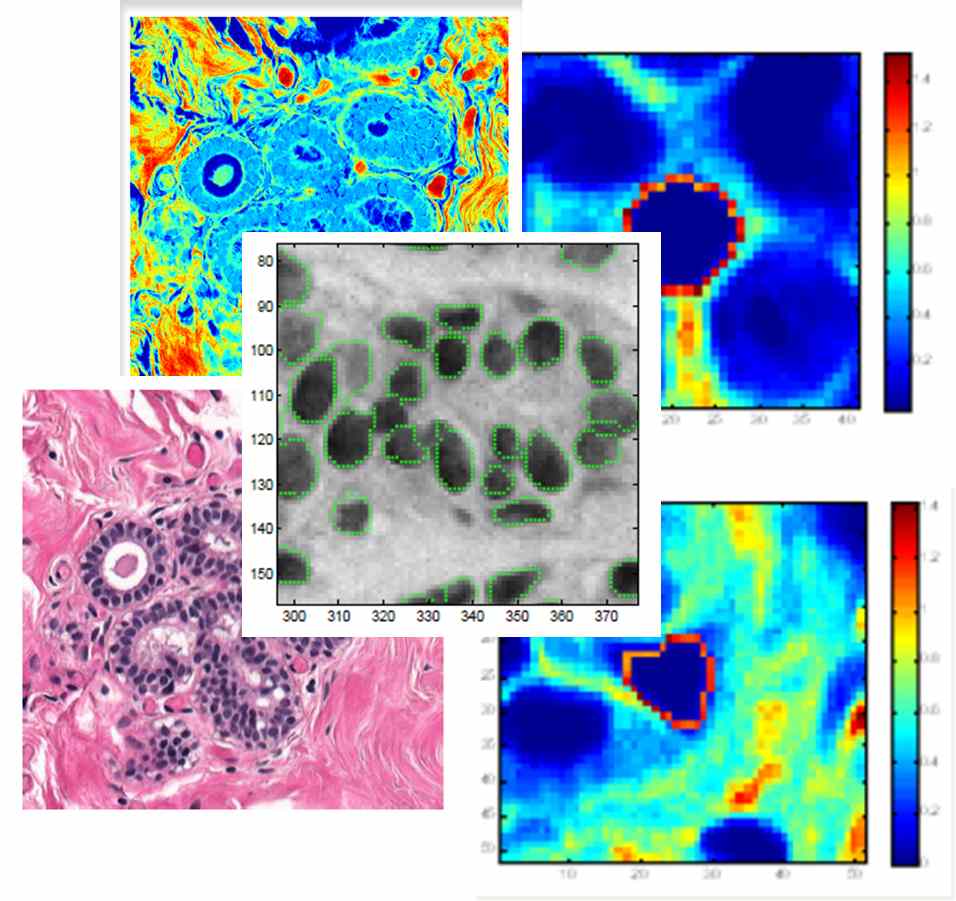
Master Thesis - Automatic Malignancy Detection in Breast Histopathology Images - Siemens Corporate Research, Princeton, NJ and TU Munich, GermanyAbstract: In traditional cancer diagnoses, pathologists examine biopsies to make diagnostic assessments largely based on deviations in the cell structures and changes in the cell distribution across the tissue. Detection of malignancy from histopathological images of breast cancer is a labor-intensive and error-prone process. Automation of this process is highly desirable and would streamline the clin- ical workflow. In this work, we present an efficient Computer Aided Diagnosis system that can differentiate between cancerous and noncancerous H&E (hemotoxylin&eosin) biopsy samples. The proposed approach aims at extracting relevant features that are used to quantify the observed changes in cancer tissue. We explore texton-based, network-based and novel morphometric features that take advantage of the special shape of the nuclei cells in breast cancer histopathological images. In extracting the network-based and morphometric features, the exact locations and segmentation of the nuclei needs to be deter- mined, which is accomplished using a generalized fast radial symmetry transform and the Random Walker algorithm, respectively. The proposed approach also uses Urquhart graph generation that can better discriminate between different anatomical structures being an approximation of the relative neighborhood graphs, which are known for matching human perceptions of the shape of a given set of points. Using a Support Vector Machine classifier on these features in conjunction with specific feature selection techniques, we differentiate between malignant and benign breast cancer histological slides. Experiments were conducted using H& E stained samples, previously annotated by pathologists. Our method achieved a high average sensitivity and specificity. |
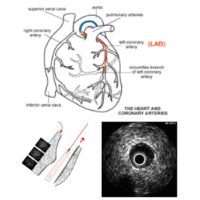
Assessment of Fluid Tissue Interaction Using Multi-Modal Image Fusion for Characterization and Progression of Coronary AtherosclerosisCoronary artery diseases such as atherosclerosis are the leading cause of death in the industrialized world. In this project, we develop computational tools for segmentation and registration problems on intravascular images including IVUS (Intravascular Ultrasound) and OCT (Optical Coherence Tomography). One sample component of this project is Automatic Stent Implant Follow-up from Intravascular OCT Pullbacks. The stents are automatically detected and their distribution is analyzed for monitoring of the stents: their malpositioning and/or tissue growth over stent struts. |
Other Work
- Bachelor's Thesis: Coding and Decoding on a Binary Transmission Channel Polytechnic University of Bucharest, Romania
Awards
- Best student finalist award at the international SPIE 2012 Medical Conference in San Diego in USA (2011)
Publications
| 2012 | |
| A. Chekkoury, Parmeshwar Khurd, Jie Ni, Claus Bahlmann, A. Khamene, Amar Patel, L. Grady, Maneesh Singh, M. Groher, N. Navab, Jeffrey Johnson, Anna Graham, Ronald Weinstein
Automated Malignancy Detection in Breast Histopathological Images SPIE Medical Imaging, 04-09 February 2012, San Diego, California, USA (bib) |
|
|
Claus Bahlmann, Amar Patel, Jeffrey Johnson, Jie Ni, A. Chekkoury, Parmeshwar Khurd, L. Grady, Anna Graham, Ronald Weinstein
Automated Detection of Diagnostically Relevant Regions in H&E Stained Digital Pathology Slides SPIE Medical Imaging, 04-09 February 2012, San Diego, California, USA (bib) |
|
| UsersForm | |
|---|---|
| Title: | |
| Firstname: | Andrei |
| Middlename: | |
| Lastname: | Chekkoury |
| Picture: | |
| Birthday: | |
| Nationality: | Romania |
| Languages: | English |
| Groups: | |
| Expertise: | |
| Position: | External Collaborator |
| Status: | Active |
| Emailbefore: | chekkour |
| Emailafter: | in.tum.de |
| Room: | |
| Telephone: | +49 89 3187 2003 |
| Alumniactivity: | |
| Defensedate: | |
| Thesistitle: | |
| Alumnihomepage: | |
| Personalvideo01: | |
| Personalvideotext01: | |
| Personalvideopreview01: | |
| Personalvideo02: | |
| Personalvideotext02: | |
| Personalvideopreview02: | |
