Beatrice Demiray geb. Lentes, M.Sc.
Contact
 |
|
Research Topics and Interests
- Multimodal Image Registration
- Medical Image Segmentation
- Deep Learning for Medical Applications
- Computer Assisted Interventions / Surgery
- Computer Aided Diagnosis
- Medical Device Tracking and Detection
- Intra-operative Navigation
- Microscopy Image Analysis
Active Research
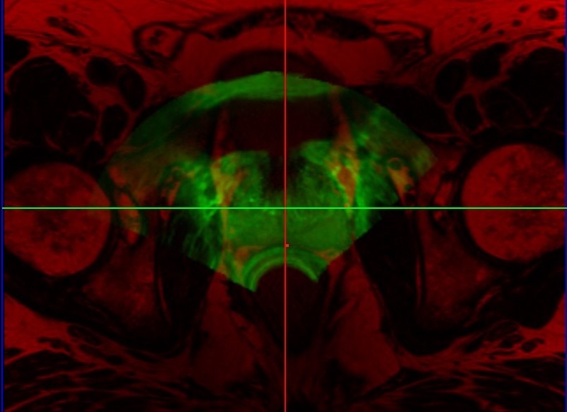
Prostate Fusion BiopsyTransrectal ultrasound (TRUS) guided biopsy remains the gold standard for diagnosis. However, it suffers from low sensitivity, leading to an elevated rate of false negative results. On the other hand, the recent advent of PET imaging using a novel dedicated radiotracer, Ga-labelled PSMA (Prostate Specific Membrane Antigen), combined with MR provides improved preinterventional identification of suspicious areas. Thus, MRI/TRUS fusion image-guided biopsy has evolved to be the method of choice to circumvent the limitations of TRUS-only biopsy. We propose a multimodal fusion image-guided biopsy framework that combines PET-MRI images with TRUS. Based on open-source software libraries, it is low cost, simple to use and has minimal overhead in clinical workflow. It is ideal as a research platform for the implementation and rapid bench to bedside translation of new image registration and visualization approaches. |
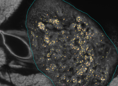
Kooperationsprojekt SFB 824 (3. Förderperiode) & BFSThe SFB824 (Sonderforschungsbereich 824: Central project for histopathology, immunohistochemistry and analytical microscopy) represents an interdisciplinary consortium which aims at the development of novel imaging technologies for the selection and monitoring of cancer therapy as an important support for personalized medicine. Z2, the central unit for comparative morphomolecular pathology and computational validation, provides integration, registration and quantification of data obtained from both macroscopic and (sub-)cellular in-vivo as well as ex-vivo imaging modalities with tissue-based morphomolecular readouts as the basis for the development and establishment of personalized medicine. In order to develop novel imaging technologies, co-annotation and validation of image data acquired by preclinical or diagnostic imaging platforms via tissue based quantitative morphomolecular methods is crucial. Light sheet microscopy will continue to close the gap between 3D data acquired by in-vivo imaging and 2D histological slices especially focusing on tumor vascularization. The Multimodal ImagiNg Data Flow StUdy Lab (MINDFUL) is a central system for data management in preclinical studies developed within SFB824. Continuing the close collaboration of pathology, computer sciences and basic as well as translational researchers from SFB824 will allow the Z2 to develop and subsequently provide a broad variety of registration and analysis tools for joint imaging and tissue based image standardization and quantification.The goal of the BFS Project: ImmunoProfiling using Neuronal Networks (IPN2) is to develop a method based on neuronal networks and recent advances in Deep Learning to allow characterization of a patient's tumor as ″hot″ or ″cold″ tumor depending on the identified ImmunoProfile. Recent research has shown that many tumors are infiltrated by immuno-competent cells, as well as that the amount, type and location of the infiltrated lymph nodes in primary tumors provide valuable prognostic information. In contrast to a ″cold tumor″, a ″hot tumor″ is characterized by an active immune system which the tumor has identified as threat. This identification provides the basis for selecting the therapy best suitable for the individual patient. |
MedInnovate: From unmet clinical needs to solution conceptsLearn how to successfully identify unmet clinical needs within the clinical routine and work towards possible and realistic solutions to solve those needs. Students will get to know tools helping them to be successful innovators in medical technology. This will include all steps from needs finding and selection to defining appropriate solution concepts, including the development of first prototypes. Get introduced to necessary steps for successful idea and concept creation and realize your project in an interdisciplinary teams comprising of physicists, informations scientists and business majors. During the project phase, you are supported by coaches from both industry and medicine, in order to allow for direct and continuous exchange. |
Teaching
Winter term 2019/20- Praktikum: Project Management and Software Development for Medical Applications (Ws 2019/20)
- Praktikum: Project Management and Software Development for Medical Applications (Ss 2019)
- Master Seminar: Lost in Translation: The Challenges of Research Transfer into Clinical Practice (Ss 2019)
- Praktikum: MedInnovate Graduate Program (Ss 2019)
- Praktikum: Project Management and Software Development for Medical Applications (Ws 2018/19)
- Praktikum: MedInnovate Graduate Program (Ws 2018/19)
- Master Seminar (Tutor): Deep Learning for Medical Applications (Ws 2018/19)
- Praktikum: Project Management and Software Development for Medical Applications (Ss 2018)
- Master Seminar: Lost in Translation: The Challenges of Research Transfer into Clinical Practice (Ss 2018)
- Master Seminar (Tutor): Deep Learning for Medical Applications (Ss 2018)
- Praktikum: Project Management and Software Development for Medical Applications (Ws 2017/18)
- Master Seminar (Tutor): Foundations of Computer Vision (Ws 2017/18)
- Master Seminar (Tutor): Deep Learning for Medical Applications (Ws 2017/18)
- Praktikum: Project Management and Software Development for Medical Applications (Ss 2017)
- Master Seminar (Tutor): Computer Vision in Animal Behaviour Studies (Ss 2017)
Student Projects
Please feel free to contact me via e-mail or drop by and ask!- Florian Albrecht: Out-of-Distribution Detection for Higher Confidence in Prostate Cancer Classification (Master Thesis Ws2020) - running
- Daniel Scherzer: Multi-structure Segmentation of Intra-Operative 3D Echocardiography Data (Master Thesis Ws2020) - running
- Christiane Weber: DigiBioP: Digital Biopsy of Prostate Lesions using PET/MR (Master Thesis Ws2020) - running
- Maria Bordukova: DigiBioP (HiWi 2020-2021) - running
- Yan-Chi Chan: DigiBioP: Database for Retrospective Prostate Cancer Study (HiWi 2019 - 2020) - finished
- Theofilos Christodolou: Vessel Quantification in MRI (IDP / Software Development Project WS2019/20) - finished
- Anna Zapaishchykova: 3D Freehand Ultrasound Spatial Calibration (IDP / Software Development Project Ss2019) - finished
- Shyam Srinivasan: Optimization of short-pulsed and high-energy laser diode driver for fast multispectral optoacoustic endoscopy (Master Thesis Ss2019) - finished
- Michael Wengler: Segmenting Blood Vessels in LSFM With Deep Neural Networks (Master Thesis Ss2019) - finished
- Muhammad Arsalan: Robust and Accurate Heart-Rate Sensing using mm-Wave Radar (Master Thesis Ss2019) - finished
- Helge Hecht: Learning Deep Similarity Metrics for Whole Slide Image Registration to Quantify Intra-Tumoural Heterogeneity (Master Thesis Ss2019) - finished
- Michael Wengler: Arivis Vision4D plugin for automatic vessel segmentation in LSFM stacks (IDP / Software Development Project Ws2018) - finished
- Khac Thanh-An Le: Blood Vessel Quantification in MRI (IDP / Software Development Project Ws2018) - finished
- Stevica Bozhinoski: Deep Learning Methods for Soft Tissue Segmentation in Ultrasound Brain Imaging (Master Thesis Ss2018) - finished
- Liesa Weigert: Deep Neural Networks for Pelvic Organ Segmentation in Multiparametic MRI (Master Thesis Ss2018) - finished
- Arsalan Muhammad: HistoTool: Advanced Annotations and Coregistration of Histological and Pre-Clinical Images (IDP / Software Development Project Ws2017) - finished
- Sergio Quijano Rojas: Development of a Medical Intervention Preparation Interface (IDP / Software Development Project Ss2017) - finished
Publications
 Scholar
| 2018 | |
| K. Westenfelder, B. Lentes, J. Rackerseder, N. Navab, J. Gschwend, M. Eiber, C. R. Maurer, Jr.
Gallium-68 HBED-CC-PSMA Positron Emission Tomography/Magnetic Resonance Imaging for Prostate Fusion Biopsy Clinical genitourinary cancer, August 2018, Volume 16, Issue 4, Pages 245-247 (bib) |
|
| B. Busam, P. Ruhkamp, S. Virga, B. Lentes, J. Rackerseder, N. Navab, C. Hennersperger
Markerless Inside-Out Tracking for 3D Ultrasound Compounding International Conference on Medical Image Computing and Computer Assisted Interventions (MICCAI), Point-of-Care Ultrasound, Granada, Spain, September 2018 [oral]. (bib) |
|
| 2017 | |
| O. Zettinig, J. Rackerseder, B. Lentes, T. Maurer, K. Westenfelder, M. Eiber, B. Frisch, N. Navab
Preconditioned Intensity-Based Prostate Registration using Statistical Deformation Models IEEE International Symposium on Biomedical Imaging (ISBI), Melbourne, April 2017. (bib) |
|


