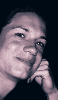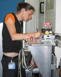
 Lorenz König taking notes of an experimental setup at ESRF on 2008-07-08.
Dipl.-Inf. Univ. Lorenz König /køːniɡ̊/
| Natural languages: | New High German (first language), Modern English, Modern French |
| Contact: | koenil01‹аτ›nyumc.org +1 (212) 26-33258 |
| Current location: | Center for Health Informatics and Bioinformatics NYU Medical Center 227 East 30th Street, 7th floor New York, NY 10016 USA |
Current Projects
Collaborative Approaches to Image Analysis
Manual and semi-automatic segmentation still playing a considerable role in medical image analysis, the collaboration of more than one human observer (segmentor) may be desirable for several reasons, including split-up of workload, review of results by a more experienced observer, and estimation of multi-observer variability. To this end, a framework is currently being designed, allowing for management of original image data as well as multiple users’ contributions, and supporting consolidation into a concensus along with an estimate of its uncertainty. A prototype of a such framework is being implemented, including basic segmentation tools, thus creating the foundations for further refinement of collaborative approaches.Analysis of Sub-Voxel Edge Localization Errors in Non-Ideal Modalities
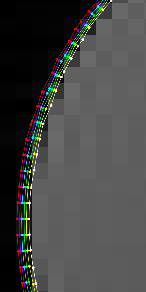
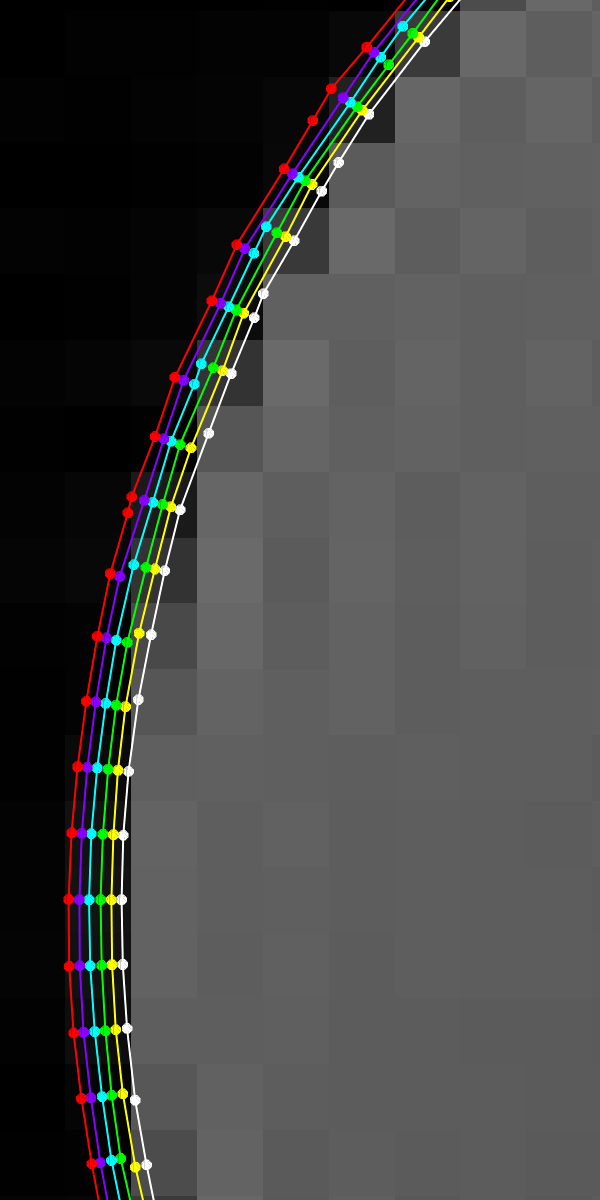
Conventional sub-voxel edge detectors act on the assumption of a box-shaped voxel aperture, or, using an equivalent formulation, point spread function (PSF). This assumtion is quite well fulfilled by industrial digital optical cameras, thus not having been an issue in the application of sub-pixel edge detectors which in the beginning was focused on aerial imaging and materials testing. However, modalities like MRI have a significantly non-box voxel aperture, a fact that results in systematic edge localization errors (which we might call “edge aberration”). First results show that by an analysis and simple correction of these errors, edge localization can be improved by about half an order of magnitude. see also: details belowDevelopment of the “PaCaSe” Software
Originating from some of my student research projects (see below), the PaCaSe software (“Patellar Cartilage Segmentation”) has been of great help to its users. It has hence been extended, now also comprising tools for thickness analysis and visualization and featuring input/output for a variety of data formats. PaCaSe will be superseded by the new collaborative framework (see above). see also: Semi-Automatic Patellar Cartilage SegmentationPublications
Student Research Projecs
Diplomarbeit “Digital Tomosynthesis in X-Ray Phase Contrast Imaging” at ESRFon the applicability of the principle and notably the algorithms of digital tomosynthesis on the emerging imaging technique of x-ray phase contrast
advisors: Alberto Bravin, Paola Coan, Tobias Lasser (2008/2009) SEP “Assessment of Knee Cartilage Thickness using Magnetic Resonance Imaging”
advisor: Ben Glocker (2007) IDP “Development and Implementation of a User-Friendly Tool for Analysis of the Patella Cartilage Using Magnetic Resonance Tomography”
advisor: Martin Groher et al. (2006/2007)
Sub-voxel Edge Detection in Non-ideal Modalities
| UsersForm | |
|---|---|
| Title: | Dipl.-Inf. Univ. |
| Firstname: | Lorenz |
| Middlename: | |
| Lastname: | König |
| Picture: | |
| Birthday: | |
| Nationality: | Germany |
| Languages: | English, German, French, Bavarian |
| Groups: | |
| Expertise: | Registration/Visualization, Segmentation, Medical Imaging, Computer Vision |
| Position: | External |
| Status: | Active |
| Emailbefore: | koenil01 |
| Emailafter: | nyumc.org |
| Room: | |
| Telephone: | |
| Alumniactivity: | |
| Defensedate: | |
| Thesistitle: | |
| Alumnihomepage: | |
| Personalvideo01: | |
| Personalvideotext01: | |
| Personalvideopreview01: | |
| Personalvideo02: | |
| Personalvideotext02: | |
| Personalvideopreview02: | |
| Circumference of the head: | |


